There is considerable inter-individual and inter-population variability in response to viruses. The potential of monocytes to elicit type-I interferon responses has attracted attention to their role in viral infections. Here, we use single-cell RNA-sequencing to characterize the role of cellular heterogeneity in human variation of monocyte responses to influenza A virus (IAV) exposure. We show widespread inter-individual variability in the percentage of IAV-infected monocytes. Notably, individuals with high cellular susceptibility to IAV are characterized by a lower activation at basal state of an IRF/STAT-induced transcriptional network, which includes antiviral genes such as IFITM3, MX1 and OAS3. Upon IAV challenge, we find that cells escaping viral infection display increased mRNA expression of type-I interferon stimulated genes and decreased expression of ribosomal genes, relative to both infected cells and those never exposed to IAV. We also uncover a stronger resistance of CD16+ monocytes to IAV infection, together with CD16+ -specific mRNA expression of IL6 and TNF in response to IAV. Finally, using flow cytometry and bulk RNA-sequencing across 200 individuals of African and European ancestry, we observe a higher number of CD16+ monocytes and lower susceptibility to IAV infection among monocytes from individuals of African-descent. Based on these data, we hypothesize that higher basal monocyte activation, driven by environmental factors and/or weak-effect genetic variants, underlies the lower cellular susceptibility to IAV infection of individuals of African ancestry relative to those of European ancestry. Further studies are now required to investigate how such cellular differences in IAV susceptibility translate into population differences in clinical outcomes and susceptibility to severe influenza.
Copyright © 2021 O’Neill, Quach, Pothlichet, Aquino, Bisiaux, Zidane, Deschamps, Libri, Hasan, Zhang, Zhang, Matuozzo, Cobat, Abel, Casanova, Naffakh, Rotival and Quintana-Murci.
Product Citations: 9
In Frontiers in Immunology on 17 December 2021 by O'Neill, M. B., Quach, H., et al.
-
FC/FACS
-
Genetics
-
Immunology and Microbiology
Quinacrine-CASIN combination overcomes chemoresistance in human acute lymphoid leukemia.
In Nature Communications on 26 November 2021 by Wu, L., Chatla, S., et al.
Chemoresistance posts a major hurdle for treatment of acute leukemia. There is increasing evidence that prolonged and intensive chemotherapy often fails to eradicate leukemic stem cells, which are protected by the bone marrow niche and can induce relapse. Thus, new therapeutic approaches to overcome chemoresistance are urgently needed. By conducting an ex vivo small molecule screen, here we have identified Quinacrine (QC) as a sensitizer for Cytarabine (AraC) in treating acute lymphoblastic leukemia (ALL). We show that QC enhances AraC-mediated killing of ALL cells, and subsequently abrogates AraC resistance both in vitro and in an ALL-xenograft model. However, while combo AraC+QC treatment prolongs the survival of primary transplanted recipients, the combination exhibits limited efficacy in secondary transplanted recipients, consistent with the survival of niche-protected leukemia stem cells. Introduction of Cdc42 Activity Specific Inhibitor, CASIN, enhances the eradication of ALL leukemia stem cells by AraC+QC and prolongs the survival of both primary and secondary transplanted recipients without affecting normal long-term human hematopoiesis. Together, our findings identify a small-molecule regimen that sensitizes AraC-mediated leukemia eradication and provide a potential therapeutic approach for better ALL treatment.
© 2021. The Author(s).
-
FC/FACS
-
Cancer Research
A bispecific CAR-T cell therapy targeting BCMA and CD38 in relapsed or refractory multiple myeloma.
In Journal of Hematology & Oncology on 9 October 2021 by Mei, H., Li, C., et al.
BCMA-specific chimeric antigen receptor-T cells (CAR-Ts) have exhibited remarkable efficacy in refractory or relapsed multiple myeloma (RRMM); however, primary resistance and relapse exist with single-target immunotherapy. Bispecific CARs are proposed to mitigate these limitations.
We constructed a humanized bispecific BM38 CAR targeting BCMA and CD38 and tested the antimyeloma activity of BM38 CAR-Ts in vitro and in vivo. Twenty-three patients with RRMM received infusions of BM38 CAR-Ts in a phase I trial.
BM38 CAR-Ts showed stronger in vitro cytotoxicity to heterogeneous MM cells than did T cells expressing an individual BCMA or CD38 CAR. BM38 CAR-Ts also exhibited potent antimyeloma activity in xenograft mouse models. In the phase I trial, cytokine release syndrome occurred in 20 patients (87%) and was mostly grade 1-2 (65%). Neurotoxicity was not observed. Hematologic toxicities were common, including neutropenia in 96% of the patients, leukopenia in 87%, anemia in 43% and thrombocytopenia in 61%. At a median follow-up of 9.0 months (range 0.5 to 18.5), 20 patients (87%) attained a clinical response and minimal residual disease-negativity (≤ 10-4 nucleated cells), with 12 (52%) achieving a stringent complete response. Extramedullary plasmacytoma was eliminated completely in 56% and partially in 33% and of 9 patients. The median progression-free survival was 17.2 months. Two relapsed patients maintained BCMA and CD38 expression on MM cells. Notably, BM38 CAR-Ts cells were detectable in 77.8% of evaluable patients at 9 months and 62.2% at 12 months.
Bispecific BM38 CAR-Ts were feasible, safe and significantly effective in patient with RRMM.
Chictr.org.cn ChiCTR1800018143.
© 2021. The Author(s).
-
FC/FACS
-
Immunology and Microbiology
In Aging (Albany NY) on 23 April 2021 by Chen, L., Guo, Z., et al.
Cancer cells-secreted extracellular vesicles (EVs) have emerged as important mediators of intercellular communication in local and distant microenvironment. Our initial GEO database analysis identified the presence of differentially-expressed microRNA-1246 (miR-1246) in acute myeloid leukemia (AML) cell-derived EVs. Consequently, the current study set out to investigate the role of AML-derived EVs-packaged miR-1246 in leukemia stem cells (LSCs) bioactivities. The predicted binding between miR-1246 and LRIG1 was verified using dual luciferase reporter assay. Then, gain- and loss-of-function assays were performed in LSCs, where LSCs were co-cultured with AML cell-derived EVs to characterize the effects of miR-1246-containing EVs, miR-1246, LRIG1 and STAT3 pathway in LSCs. Our findings revealed, in AML cell-derived EVs, miR-1246 was highly-expressed and directly-targeted LRIG1 to activate the STAT3 pathway. MiR-1246 inhibitor or EV-encapsulated miR-1246 inhibitor was found to suppress the viability and colony formation abilities but promoted the apoptosis and differentiation of LSCs through inactivation of STAT3 pathway by up-regulating LRIG1. In addition, the inhibitory effects of AML cell-derived EVs carrying miR-1246 inhibitor on LSCs were substantiated by in vivo experiments. Collectively, our findings reveal that the repression of AML cell-derived EVs containing miR-1246 inhibitor alters the survival of LSCs by inactivating the LRIG1-mediated STAT3 pathway.
-
FC/FACS
-
Cancer Research
-
Stem Cells and Developmental Biology
High CD45 expression of CD8+ and CD4+ T cells correlates with the size of HIV-1 reservoir in blood.
In Scientific Reports on 24 November 2020 by Petkov, S., Bekele, Y., et al.
Using mass cytometry, we investigated the expression of 28 markers on CD8+ and CD4+ T cells from HIV-1 infected patients with a variable size of HIV-1 reservoir defined as high (HR) and low (LR) reservoir; we aimed at identifying phenotypic associations of T cells with size of HIV-1 reservoir. We showed that the frequency of CD45+ CD8+ and CD4+ T cells was directly proportional to the size of HIV-1 reservoir; HR patients had a significantly larger frequency of blood CD45high T cells and higher CD45 expression on both CD8+ and CD4+ T cells. CD45 is a receptor-type protein tyrosine phosphatase essential in TCR signaling. Functional and phenotypical analysis of CD45high cells revealed that they express activation and proliferation markers (CD38 + HLA-DR + and Ki-67) and produce cytokines upon in vitro activation. CD45high T cells also expressed high levels of immune check-point PD-1. Our results link CD45 expression on T cells to HIV-1 reservoir; PD-1 expression on CD45high T cells may contribute to their exhaustion.
-
FC/FACS
-
Cardiovascular biology
-
Immunology and Microbiology
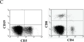
In BMC Cancer on 7 June 2011 by Lee, Y. S., Kim, T. S., et al.
Fig.1.C

-
FC/FACS
-
Collected and cropped from BMC Cancer by CiteAb, provided under a CC-BY license
Image 1 of 2
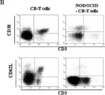
In BMC Cancer on 7 June 2011 by Lee, Y. S., Kim, T. S., et al.
Fig.2.B

-
FC/FACS
-
Collected and cropped from BMC Cancer by CiteAb, provided under a CC-BY license
Image 1 of 2