Terminal erythropoiesis is a complex multistep process involving coordination of gene transcription and dramatic nuclear condensation, which leads to the expulsion of nuclei to generate reticulocytes. However, we lack a comprehensive understanding of the key transcriptional and epigenetic regulators involved.
We used a high-throughput small molecule screen in primary CD34+-derived human erythroblasts to identify targets that promoted terminal erythropoiesis, and further confirmed the phenotype in different differentiation systems by inhibitors and shRNAs of different BRD4 isoforms. Then we performed RNA-seq, ATAC-seq, ChIP-qPCR, Co-IP, and reanalyzed previously-published transcriptional data and mass spectrometric data to clarify how BRD4 regulates terminal erythropoiesis.
We identified that inhibitors of the bromodomain protein BRD4, an epigenetic reader and transcriptional activator together with CDK9, promoted terminal erythropoiesis from hematopoietic stem/progenitor cells and embryonic stem cells, and enhanced enucleation. Combined analysis of our RNA-seq, ATAC-seq, and previously-published transcriptional data of erythroblast differentiation at different stages confirmed that BRD4 inhibition accelerates erythroblast maturation. Unexpectedly, this BRD4 function was independent of its classical CDK9 interaction and transcriptional activation. Instead, RNA-seq, ATAC-seq, and Cut&Tag upon BRD4 inhibition revealed that BRD4 regulates erythropoiesis by inhibiting the small G protein RhoB and disrupts actin reorganization. ChIP-qPCR, Co-IP, and functional studies revealed that BRD4 acts as a transcriptional repressor by interacting with the histone methyltransferase EHMT1/2.
We demonstrate a non-classical role for BRD4 as a transcriptional repressor of RhoB to regulate erythroid maturation, and classical CDK9 dependent role to regulate cell proliferation of erythroblasts. Besides, we clarify RhoB's activity and function during terminal erythropoiesis. BRD4 inhibition might be a simple method to promote in vitro blood cell production, and a candidate therapeutic target for diseases leading to dyserythropoiesis such as myelodysplastic syndromes.
© 2025. The Author(s).
Product Citations: 48
BRD4 acts as a transcriptional repressor of RhoB to inhibit terminal erythropoiesis.
In Journal of Hematology & Oncology on 1 July 2025 by Chen, Y., Huo, D., et al.
-
FC/FACS
-
Biochemistry and Molecular biology
In Stem Cells Translational Medicine on 25 June 2025 by Lennon, M. L., Frieman, A., et al.
Amniotic fluid is a promising source of autologous cells for disease modeling, drug screening, and regenerative medicine applications. However, current methods of collecting amniotic fluid are invasive, and samples are limited to pregnancies that require amniocentesis or cesarean section.
The purpose of this study was to determine whether amniotic fluid cells could be isolated and cultured from amniotic fluid collected during vaginal deliveries.
Amniotic fluid samples were obtained during delivery of 4 neonates, 3 of which had been prenatally diagnosed with hypoplastic left heart syndrome (HLHS) in utero. Adherent amniotic fluid cells were assessed for maternal cell contamination, proliferation rate, surface marker expression, and differentiation potential. Amniotic fluid cells were also reprogrammed to induced pluripotent stem cells (iPSCs) and differentiated into functional cardiomyocytes.
Amniotic fluid cells collected from vaginal deliveries showed similar surface marker phenotype and differentiation characteristics to amniotic fluid-derived mesenchymal stem cells collected from amniocentesis and cesarean section. Amniotic fluid cells collected during vaginal births of both neonates with HLHS and one neonate with typical heart geometry could be reprogrammed to iPSCs and differentiated to a cardiac lineage with high efficiency. Conclusions and Relevence: These findings suggest that amniotic fluid collected from vaginal births is a readily available source of patient-specific stem cells for banking, in vitro disease modeling, and regenerative medicine applications.
© The Author(s) 2025. Published by Oxford University Press.
-
Stem Cells and Developmental Biology
In International Journal of Stem Cells on 30 May 2025 by Lee, N. K., Na, D. L., et al.
Mesenchymal stem cells (MSCs) are frequently used for therapeutic applications in both pre-clinical and clinical settings owing to their capacity for immune modulation and neuroprotective effects. However, transient fever is commonly observed as an adverse event following MSC injection in patients with Alzheimer's disease (AD). In this study, we investigated the potential impact of immunosuppressants such as dexamethasone and tacrolimus on altering the characteristics of human mesenchymal stem cells (hMSCs). Additionally, we examined whether these immunosuppressants affect the persistence of hMSCs or the immune response upon their administration into the brain parenchyma of AD mice. The exposure of hMSCs to high concentrations of dexamethasone and tacrolimus in vitro did not significantly alter the characteristics of hMSCs. The expression of genes related to innate immune responses, such as Irak1, Irf3, Nod1, and Ifnar1, was significantly downregulated by the additional administration of dexamethasone and tacrolimus to the brain parenchyma of AD mice. However, hMSC persistence in the AD mouse brain was not affected. The results of this study support the use of immunosuppressants to mitigate fever during stem cell therapy in patients with AD.
-
FC/FACS
-
Stem Cells and Developmental Biology
Simple Isolation of Human Bone Marrow Adipose Tissue-Derived Mesenchymal Stem/Stromal Cells.
In Current Protocols on 1 January 2025 by Tonyalı, G., Kılıc, E., et al.
Bone marrow adipose tissue (BMAT) has garnered significant attention due to its critical roles in leukemia pathogenesis, cancer metastasis, and bone marrow failure. BMAT is a metabolically active, distinct tissue that differs from other fat depots. Marrow adipocytes, closely interacting with hematopoietic stem/progenitor cells and osteoblasts, play a pivotal role in regulating their functions. However, standardized methods for isolating and defining human BMAT (hBMAT) remain unclear. In animal models, BMAT is commonly isolated directly from the bone marrow through flushing, enzymatic digestion, or mechanical disruption. In humans, BMAT isolation often involves the adipogenic induction of bone marrow mesenchymal stem/stromal cells (BM-MSCs) derived from bone marrow aspirates. However, this approach reflects cellular responses to chemical stimuli and may not accurately represent in vivo differentiation potential. Similarly, BMAT obtained from hip or knee replacement surgeries might not reflect basal physiological conditions due to inflammatory influences. Here, we describe a direct method for culturing BMAT from the fatty layer of bone marrow aspirates obtained from healthy transplant donors. This protocol employs centrifugation and washing steps using basic laboratory equipment, offering simple and non-enzymatic approach. For validation, isolated cells are characterized according to the International Society for Cell & Gene Therapy (ISCT) criteria. © 2025 Wiley Periodicals LLC. Basic Protocol 1: Isolation of human BMAT-MSCs from the fatty layer of the bone marrow Basic Protocol 2: Culture expansion, trypsinization, and cryopreservation of BMAT-MSCs Support Protocol 1: Immunophenoypic characterization of human BMAT-MSCs by flow cytometry Support Protocol 2: In vitro characterization of multilineage differentiation potential of human BMAT-MSCs Support Protocol 3: Further characterization of gene expression in human BMAT-MSCs using qRT-PCR.
© 2025 Wiley Periodicals LLC.
In Stem Cell Research & Therapy on 12 October 2024 by Czosseck, A., Chen, M. M., et al.
Cell therapy can protect cardiomyocytes from hypoxia, primarily via paracrine secretions, including extracellular vesicles (EVs). Since EVs fulfil specific biological functions based on their cellular origin, we hypothesised that EVs from human cardiac stromal cells (CMSCLCs) obtained from coronary artery bypass surgery may have cardioprotective properties.
This study characterises CMSCLC EVs (C_EVs), miRNA cargo, cardioprotective efficacy and transcriptomic modulation of hypoxic human induced pluripotent stem cell-derived cardiomyocytes (iPSC-CMs). C_EVs are compared to bone marrow mesenchymal stromal cell EVs (B_EVs) which are a known therapeutic EV type.
Cells were characterised for surface markers, gene expression and differentiation potential. EVs were compared for yield, phenotype, and ability to protect hiPSC-CMs from hypoxia/reoxygenation injury. EV dose was normalised by both protein concentration and particle count, allowing direct comparison. C_EV and B_EV miRNA cargo was profiled and RNA-seq was performed on EV-treated hypoxic hiPSC-CMs, then data were integrated by multi-omics. Confirmatory experiments were carried out using miRNA mimics.
At the same dose, C_EVs were more effective than B_EVs at protecting CM integrity, reducing apoptotic markers, and cell death during hypoxia. While C_EVs and B_EVs shared 70-77% similarity in miRNA content, C_EVs contained unique miRNAs, including miR-202-5p, miR-451a and miR-142-3p. Delivering miRNA mimics confirmed that miR-1260a and miR-202/451a/142 were cardioprotective, and the latter upregulated protective pathways similar to whole C_EVs.
This study demonstrates the potential of cardiac tissues, routinely discarded following surgery, as a valuable source of EVs for myocardial infarction therapy. We also identify miR-1260a as protective of CM hypoxia.
© 2024. The Author(s).
-
Homo sapiens (Human)
-
Cardiovascular biology
-
Stem Cells and Developmental Biology
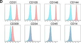
In Stem Cell Res Ther on 22 January 2019 by Gao, K., Kumar, P., et al.
Fig.1.D

-
FC/FACS
-
Homo sapiens (Human)
Collected and cropped from Stem Cell Res Ther by CiteAb, provided under a CC-BY license
Image 1 of 1