Colchicine is a broad-acting anti-inflammatory agent that has attracted interest for repurposing in atherosclerotic cardiovascular disease. Here, we studied its ability at a human equivalent dose of 0.5 mg/day to modify plaque formation and composition in murine atherosclerosis and investigated its actions on macrophage responses to atherogenic stimuli in vitro. In atherosclerosis induced by high-cholesterol diet, Apoe-/- mice treated with colchicine had 50% reduction in aortic oil Red O+ plaque area compared to saline control (p = .001) and lower oil Red O+ staining of aortic sinus lesions (p = .03). In vitro, addition of 10 nM colchicine inhibited foam cell formation from murine and human macrophages after treatment with oxidized LDL (ox-LDL). Mechanistically, colchicine downregulated glycosylation and surface expression of the ox-LDL uptake receptor, CD36, and reduced CD36+ staining in aortic sinus plaques. It also decreased macrophage uptake of cholesterol crystals, resulting in lower intracellular lysosomal activity, inhibition of the NLRP3 inflammasome, and reduced secretion of IL-1β and IL-18. Colchicine's anti-atherosclerotic actions were accentuated in a mouse model of unstable plaque induced by carotid artery tandem stenosis surgery, where it decreased lesion size by 48% (p = .01), reduced lipid (p = .006) and necrotic core area (p = .007), increased collagen content and cap-to-necrotic core ratio (p = .05), and attenuated plaque neutrophil extracellular traps (p < .001). At low dose, colchicine's effects were not accompanied by the evidence of microtubule depolymerization. Together, these results show that colchicine exerts anti-atherosclerotic and plaque-stabilizing effects at low dose by inhibiting foam cell formation and cholesterol crystal-induced inflammation. This provides a new framework to support its repurposing for atherosclerotic cardiovascular disease.
© 2023 Federation of American Societies for Experimental Biology.
Product Citations: 12
In The FASEB Journal on 1 April 2023 by Schwarz, N., Fernando, S., et al.
-
FC/FACS
-
Mus musculus (House mouse)
In HemaSphere on 1 February 2023 by Ernst, M. P. T., Pronk, E., et al.
RUNX1 familial platelet disorder (RUNX1-FPD) is a hematopoietic disorder caused by germline loss-of-function mutations in the RUNX1 gene and characterized by thrombocytopathy, thrombocytopenia, and an increased risk of developing hematologic malignancies, mostly of myeloid origin. Disease pathophysiology has remained incompletely understood, in part because of a shortage of in vivo models recapitulating the germline RUNX1 loss of function found in humans, precluding the study of potential contributions of non-hematopoietic cells to disease pathogenesis. Here, we studied mice harboring a germline hypomorphic mutation of one Runx1 allele with a loss-of-function mutation in the other Runx1 allele (Runx1 L148A/- mice), which display many hematologic characteristics found in human RUNX1-FPD patients. Runx1 L148A/- mice displayed robust and pronounced thrombocytopenia and myeloid-biased hematopoiesis, associated with an HSC intrinsic reconstitution defect in lymphopoiesis and expansion of myeloid progenitor cell pools. We demonstrate that specific deletion of Runx1 from bone marrow stromal cells in Prrx1-cre;Runx1 fl/fl mice did not recapitulate these abnormalities, indicating that the hematopoietic abnormalities are intrinsic to the hematopoietic lineage, and arguing against a driving role of the bone marrow microenvironment. In conclusion, we report a RUNX1-FPD mouse model faithfully recapitulating key characteristics of human disease. Findings do not support a driving role of ancillary, non-hematopoietic cells in the disruption of hematopoiesis under homeostatic conditions.
Copyright © 2023 the Author(s). Published by Wolters Kluwer Health, Inc. on behalf of the European Hematology Association.
-
FC/FACS
-
Mus musculus (House mouse)
In Stem Cells on 30 January 2023 by Sim, H. J., Bhattarai, G., et al.
While supplemental angiopoietin-1 (Ang1) improves hematopoiesis, excessive Ang1 induces bone marrow (BM) impairment, hematopoietic stem cell (HSC) senescence, and erythropoietic defect. Here, we examined how excessive Ang1 disturbs hematopoiesis and explored whether hematopoietic defects were related to its level using K14-Cre;c-Ang1 and Col2.3-Cre;c-Ang1 transgenic mice that systemically and locally overexpress cartilage oligomeric matrix protein-Ang1, respectively. We also investigated the impacts of Tie2 inhibitor and AMD3100 on hematopoietic development. Transgenic mice exhibited excessive angiogenic phenotypes, but K14-Cre;c-Ang1 mice showed more severe defects in growth and life span with higher presence of Ang1 compared with Col2.3-Cre;c-Ang1 mice. Dissimilar to K14-Cre;c-Ang1 mice, Col2.3-Cre;c-Ang1 mice did not show impaired BM retention or senescence of HSCs, erythropoietic defect, or disruption of the stromal cell-derived factor 1 (SDF-1)/CXCR4 axis. However, these mice exhibited a defect in platelet production depending on the expression of Tie2 and globin transcription factor 1 (GATA-1), but not GATA-2, in megakaryocyte progenitor (MP) cells. Treatment with Tie2 inhibitor recovered GATA-1 expression in MP cells and platelet production without changes in circulating RBC in transgenic mice. Consecutive AMD3100 administration not only induced irrecoverable senescence of HSCs but also suppressed formation of RBC, but not platelets, via correlated decreases in number of erythroblasts and their GATA-1 expression in B6 mice. Our results indicate that genetic overexpression of Ang1 impairs hematopoietic development depending on its level, in which megakaryopoiesis is preferentially impaired via activation of Ang1/Tie2 signaling, whereas erythropoietic defect is orchestrated by HSC senescence, inflammation, and disruption of the SDF-1/CXCR4 axis.
© The Author(s) 2022. Published by Oxford University Press.
-
FC/FACS
-
Stem Cells and Developmental Biology
Chronic allergic lung inflammation negatively influences neurobehavioral outcomes in mice.
In Journal of Neuroinflammation on 31 August 2022 by Kanaya, A., Yang, M., et al.
Asthma is a major public health problem worldwide. Emerging data from epidemiological studies show that allergies and allergic diseases may be linked to anxiety, depression and cognitive decline. However, little is known about the effect of asthma, an allergic lung inflammation, on cognitive decline/behavioral changes. Therefore, we investigated the hypothesis that allergic lung inflammation causes inflammation in the brain and leads to neurobehavioral changes in mice.
Wild-type C57BL/6J female mice were sensitized with nasal house dust mite (HDM) antigen or control PBS for 6 weeks to induce chronic allergic lung inflammation. A series of neurocognitive tests for anxiety and/or depression were performed before and after the intranasal HDM administration. After the behavior tests, tissues were harvested to measure inflammation in the lungs and the brains.
HDM-treated mice exhibited significantly increased immobility times during tail suspension tests and significantly decreased sucrose preference compared with PBS controls, suggesting a more depressed and anhedonia phenotype. Spatial memory impairment was also observed in HDM-treated mice when assessed by the Y-maze novel arm tests. Development of lung inflammation after 6 weeks of HDM administration was confirmed by histology, bronchoalveolar lavage (BAL) cell count and lung cytokine measurements. Serum pro-inflammatory cytokines and Th2-related cytokines levels were elevated in HDM-sensitized mice. In the brain, the chemokine fractalkine was increased in the HDM group. The c-Fos protein, a marker for neuronal activity, Glial Fibrillary Acidic Protein (GFAP) and chymase, a serine protease from mast cells, were increased in the brains from mice in HDM group. Chymase expression in the brain was negatively correlated with the results of sucrose preference rate in individual mice.
6 weeks of intranasal HDM administration in mice to mimic the chronic status of lung inflammation in asthma, caused significant inflammatory histological changes in the lungs, and several behavioral changes consistent with depression and altered spatial memory. Chymase and c-Fos proteins were increased in the brain from HDM-treated mice, suggesting links between lung inflammation and brain mast cell activation, which could be responsible for depression-like behavior.
© 2022. The Author(s).
-
FC/FACS
-
Mus musculus (House mouse)
-
Immunology and Microbiology
Mds1CreERT2, an inducible Cre allele specific to adult-repopulating hematopoietic stem cells.
In Cell Reports on 17 August 2021 by Zhang, Y., McGrath, K. E., et al.
Hematopoietic ontogeny consists of two broad programs: an initial hematopoietic stem cell (HSC)-independent program followed by HSC-dependent hematopoiesis that sequentially seed the fetal liver and generate blood cells. However, the transition from HSC-independent to HSC-derived hematopoiesis remains poorly characterized. To help resolve this question, we developed Mds1CreERT2 mice, which inducibly express Cre-recombinase in emerging HSCs in the aorta and label long-term adult HSCs, but not HSC-independent yolk-sac-derived primitive or definitive erythromyeloid (EMP) hematopoiesis. Our lineage-tracing studies indicate that HSC-derived erythroid, myeloid, and lymphoid progeny significantly expand in the liver and blood stream between E14.5 and E16.5. Additionally, we find that HSCs contribute the majority of F4/80+ macrophages in adult spleen and marrow, in contrast to their limited contribution to macrophage populations in brain, liver, and lungs. The Mds1CreERT2 mouse model will be useful to deconvolute the complexity of hematopoiesis as it unfolds in the embryo and functions postnatally.Copyright © 2021. Published by Elsevier Inc.
-
Mus musculus (House mouse)
-
Stem Cells and Developmental Biology
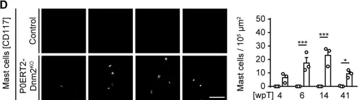
In Elife on 16 January 2019 by Gerber, D., Ghidinelli, M., et al.
Fig.3.D

-
ICC
-
Mus musculus (House mouse)
Collected and cropped from Elife by CiteAb, provided under a CC-BY license
Image 1 of 1