Abstract Background. X-linked retinoschisis is a retinovitreal disorder primarily affecting males, starting in childhood. Over time, patients experience deterioration of vision due to the lack of retinoschisin-1 function. In clinical trials performing ocular gene delivery in those affected by this disorder, ocular inflammation was observed, which may have masked efficacy. A subsequent study focusing on analyzing the populations of peripheral blood mononuclear cells and cytokines in adults with this disease reported aberrant dendritic cell numbers and cytokine levels in peripheral blood. Adults with this disease may have an altered baseline immunity. However, children with X-linked retinoschisis were not included in that study, and whether the aberrant peripheral immunity in affected adults was a consequence of advanced eye pathology remained unclear. This study focuses on analyzing the populations of blood lymphocyte subsets in children aged 0 to 7 with X-linked retinoschisis and age-matched controls using flow cytometry. Results. The fractions of lymphocyte subsets that were CD16a + were significantly lower in children with X-linked retinoschisis. The fractions of lymphocyte subsets that were CD16a+/CD56+, namely natural killer cells, were also significantly lower. In children with X-linked retinoschisis, the fractions of CD3+/CD4 + T cells were higher, and the fractions of CD3 + CD8 + T cells were lower, despite having the same amounts of total CD3 + T cells within their lymphocyte populations. This resulted in a significantly greater CD4/CD8 ratio in children with X-linked retinoschisis compared to age-matched controls. Conclusions. Alterations were found in blood lymphocyte compositions of children with X-linked retinoschisis within both innate and adaptive immune axes. Some alterations including an elevation of CD4/CD8 ratio in X-linked retinoschisis mirror those previously found in adult patients with this disease. The fact that these abnormalities were present early in this disease indicates that retinoschisin-1 may play a role in regulating immunity in addition to retinal adhesion. The findings may have implications for future treatments such as ocular gene delivery.
Product Citations: 27
Immune landscape in children with X-linked retinoschisis
Preprint on Research Square on 17 April 2025 by Hsu, Y., Valle, G. D., et al.
-
Immunology and Microbiology
In Cancers on 7 September 2024 by Alaklabi, S., Maguire, O., et al.
The CLARINET trial led to the approval of lanreotide for the treatment of patients with gastroenteropancreatic neuroendocrine tumors (NETs). It is hypothesized that lanreotide regulates proliferation, hormone synthesis, and other cellular functions via binding to somatostatin receptors (SSTR1-5) present in NETs. However, our knowledge of how lanreotide affects the immune system is limited. In vitro studies have investigated functional immune response parameters with lanreotide treatment in healthy donor T cell subsets, encompassing the breadth of SSTR expression, apoptosis induction, cytokine production, and activity of transcription factor signaling pathways. In our study, we characterized in vitro immune mechanisms in healthy donor T cells in response to lanreotide. We also studied the in vivo effects by looking at differential gene expression pre- and post-lanreotide therapy in patients with NET. Immune-focused gene and protein expression profiling was performed on peripheral blood samples from 17 NET patients and correlated with clinical response. In vivo, lanreotide therapy showed reduced effects on wnt, T cell receptor (TCR), and nuclear factor kappa-light-chain-enhancer of activated B cells (NF-kB) signaling in CD8+ T cells in responders compared to non-responders. Compared to non-responders, responders showed reduced effects on cytokine and chemokine signaling but greater effects on ubiquitination and proteasome degradation genes. Our results suggest significant lanreotide pharmacodynamic effects on immune function in vivo, which correlate with responses in NET patients. This is not evident from experimental in vitro settings.
-
Cancer Research
-
Endocrinology and Physiology
-
Immunology and Microbiology
In Nature Communications on 14 August 2024 by Münchhalfen, M., Goerg, R., et al.
Ligation of the B cell antigen receptor (BCR) initiates humoral immunity. However, BCR signaling without appropriate co-stimulation commits B cells to death rather than to differentiation into immune effector cells. How BCR activation depletes potentially autoreactive B cells while simultaneously primes for receiving rescue and differentiation signals from cognate T lymphocytes remains unknown. Here, we use a mass spectrometry-based proteomic approach to identify cytosolic/nuclear shuttling elements and uncover transcription factor EB (TFEB) as a central BCR-controlled rheostat that drives activation-induced apoptosis, and concurrently promotes the reception of co-stimulatory rescue signals by supporting B cell migration and antigen presentation. CD40 co-stimulation prevents TFEB-driven cell death, while enhancing and prolonging TFEB's nuclear residency, which hallmarks antigenic experience also of memory B cells. In mice, TFEB shapes the transcriptional landscape of germinal center B cells. Within the germinal center, TFEB facilitates the dark zone entry of light-zone-residing centrocytes through regulation of chemokine receptors and, by balancing the expression of Bcl-2/BH3-only family members, integrates antigen-induced apoptosis with T cell-provided CD40 survival signals. Thus, TFEB reprograms antigen-primed germinal center B cells for cell fate decisions.
© 2024. The Author(s).
-
FC/FACS
-
Mus musculus (House mouse)
-
Homo sapiens (Human)
-
Immunology and Microbiology
In American Journal of Reproductive Immunology (New York, N.Y. : 1989) on 1 June 2023 by Muyayalo, K. P., Song, S., et al.
This study aimed to identify subsets of regulatory T cells (Tregs) associated with ovarian aging and determine whether they can be used as markers of reproductive aging.
This prospective cohort study was conducted among women of reproductive age. Basic physiological characteristics, reproductive hormones, Treg cell subsets, and correlations between these parameters were assessed. The POSEIDON criteria was used to identify women with low reproductive potential.
The percentages of HLA-DR+ CD45RA- Tregs and CD28- Treg-like cells significantly increased with age. Women between 40 and 49 years had significantly higher percentages of HLA-DR+ CD45RA- Tregs and CD28- Treg-like cells than those at 20-29, 30-34, and 35-39 years old. Age positively correlated with FSH levels and the percentages of HLA-DR+ CD45RA- Tregs and CD28- Treg-like cells, but inversely correlated with antral follicle count (AFC) and AMH levels. Interestingly, a positive correlation was found between the percentages of HLA-DR+ CD45RA- Tregs and FSH levels, whereas an inverse correlation was found between those of HLA-DR+ CD45RA- Tregs and AFC or AMH levels. Furthermore, a significant positive correlation was observed between the percentages of CD28- Treg-like cells and AFC. Based on POSEIDON criteria, women with the percentages of HLA-DR+ CD45RA- Tregs and CD28- Treg-like cells above reference value ranges were assigned to the low prognosis groups.
These findings suggest that HLA-DR+ CD45RA- Tregs and CD28- Treg-like cells can be used as immunologic markers of reproductive aging, which helps clinicians identify women with low reproductive potential and establish individualized therapeutic strategies.
© 2022 John Wiley & Sons A/S. Published by John Wiley & Sons Ltd.
-
Immunology and Microbiology
Do more with Less: Improving High Parameter Cytometry Through Overnight Staining.
In Current Protocols on 1 November 2022 by Whyte, C. E., Tumes, D. J., et al.
Recent advances in flow cytometry have allowed high-dimensional characterization of biological phenomena, enabling breakthroughs in a multitude of fields. Despite the appreciation of the unique properties of antigens and fluorophores in high-parameter panel design, staining conditions are often standardized for short surface stains, regardless of antibody affinity or antigen accessibility. Here, we demonstrate how increasing antibody incubation times can lead to substantial improvements in sensitivity, maintaining specificity, and reducing background, while also significantly reducing the costs of high-parameter cytometry panels. Furthermore, overnight staining reduces the influence of interexperimental variability, assisting accurate pooling over experiments over extended time courses. We provide guidance on how to optimize staining conditions for diverse antigens, including how different fixation strategies can affect epitope accessibility. Overnight staining can thus substantially improve the resolution, repeatability, and cost-effectiveness of high-parameter cytometry. © 2022 The Authors. Current Protocols published by Wiley Periodicals LLC.
© 2022 The Authors. Current Protocols published by Wiley Periodicals LLC.
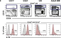
In Dev Cell on 5 February 2018 by Böiers, C., Richardson, S. E., et al.
Fig.1.B

-
FC/FACS
-
Collected and cropped from Dev Cell by CiteAb, provided under a CC-BY license
Image 1 of 1