Limited therapeutic options are available for patients with breast cancer brain metastases (BCBM), and thus there is an urgent need for novel treatment approaches. We previously engineered an effective oncolytic herpes simplex virus 1 (oHSV) expressing a full-length anti-CD47 monoclonal antibody (mAb) with a human IgG1 scaffold (OV-αCD47-G1) that was used to treat both ovarian cancer and glioblastoma. Here, we demonstrate that the combination of OV-αCD47-G1 and temozolomide (TMZ) improve outcomes in preclinical models of BCBM. The combination of TMZ with OV-αCD47-G1 synergistically increased macrophage phagocytosis against breast tumor cells and led to greater activation of NK cell cytotoxicity. In addition, the combination of OV-αCD47-G1 with TMZ significantly prolonged the survival of tumor-bearing mice when compared with TMZ or OV-αCD47-G1 alone. Combination treatment with the mouse counterpart of OV-αCD47-G1, termed OV-A4-IgG2b, also enhanced mouse macrophage phagocytosis, NK cell cytotoxicity, and survival in an immunocompetent model of mice bearing BCBM compared with TMZ or OV-A4-IgG2b alone. Collectively, these results suggest that OV-αCD47-G1 combined with TMZ should be explored in patients with BCBM.
© 2024 The Authors. Published by Elsevier Inc. on behalf of The American Society of Gene and Cell Therapy.
Product Citations: 9
In Molecular Therapy. Oncology on 19 September 2024 by Wang, J., Tian, L., et al.
-
Cancer Research
-
Immunology and Microbiology
In Scientific Reports on 10 September 2024 by Hansen, F. J., Mittelstädt, A., et al.
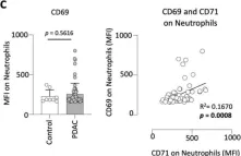
Pancreatic ductal adenocarcinoma (PDAC) is one of the most lethal malignancies, presenting a persisting global health burden. Neutrophils have a double-edged role in tumor progression exhibiting both pro-tumor and anti-tumor functions. CD71, also known as transferrin receptor 1, performs a critical role in cellular iron uptake and is highly expressed on proliferating cells, and especially on activated immune cells. CD71 is known to be elevated in various types of solid cancers and is associated with poor prognosis, however, the expression of CD71 on neutrophils in PDAC and its potential clinical impact is still unknown. Therefore, we analyzed CD71 on circulating neutrophils in PDAC and clinical control patients and found a significant increased expression in PDAC patients. High expression of CD71 on neutrophils in PDAC patients was associated with reduced outcome compared to low expression. CD71 on neutrophils correlated positively with the levels of proinflammatory cytokines IL-6, IFN-γ, and growth factor ligands CD40-L, and BAFF in plasma of PDAC patients. Finally, we have demonstrated that high expression of CD71 on neutrophils was also associated with an increased expression of CD39 and CD25 on circulating T-cells. Based on our findings, we hypothesize that CD71 on neutrophils is associated with tumor progression in PDAC. Further studies are required to investigate the distinct functionality of CD71 expressing neutrophils and their potential clinical application.
© 2024. The Author(s).
-
FC/FACS
-
Homo sapiens (Human)
-
Cancer Research
In STAR Protocols on 15 March 2024 by Gail, D. P., Suzart, V. G., et al.
M1- and M2-like macrophages infected with Mycobacterium tuberculosis (Mtb) have been found to differ in their capacity to elicit memory CD4+ T cell activation. Here, we present a protocol to quantify and isolate the subset of human memory CD4+ T cells activated in response to autologous monocyte-derived macrophages (MDMs) infected with virulent Mtb. We describe steps for CD14+ monocyte isolation, generating MDMs, culturing Mtb and infection of macrophages, and identifying activated CD4+ T cells by flow cytometry. For complete details on the use and execution of this protocol, please refer to Gail et al.1.
Copyright © 2024 The Author(s). Published by Elsevier Inc. All rights reserved.
-
Immunology and Microbiology
In Frontiers in Immunology on 19 January 2024 by The, S. M. L., Schreurs, R. R. C. E., et al.
Appendicitis is one of the most common causes of acute abdominal surgery in children. The clinical course of appendicitis ranges from simple to complex appendicitis. The mechanisms underlying the heterogeneity of appendicitis in children remain largely unclear. Dysregulated T cell responses play an important role in several inflammatory diseases of the intestine, but the extend of T cell dysregulation in appendicitis in children is less well known.
To characterize appendiceal T cells in simple and complex appendicitis we performed in-depth immunophenotyping of appendiceal-derived T cells by flow cytometry and correlated this to appendiceal-derived microbiota analyses of the same patient.
Appendix samples of twenty children with appendicitis (n = 8 simple, n = 12 complex) were collected. T cells in complex appendicitis displayed an increased differentiated phenotype compared to simple appendicitis, including a loss of both CD27 and CD28 by CD4+ T cells and to a lesser extent by CD8+ T cells. Frequencies of phenotypic tissue-resident memory CD69+CD4+ T cells and CD69+CD8+ T cells were decreased in children with complex compared to simple appendicitis, indicating disruption of local tissue-resident immune responses. In line with the increased differentiated phenotype, cytokine production of in particular IL-17A by CD4+ T cells was increased in children with complex compared to simple appendicitis. Furthermore, frequencies of IL-17A+ CD4+ T cells correlated with a dysregulation of the appendiceal microbiota in children with complex appendicitis.
In conclusion, disruption of local T cell responses, and enhanced pro-inflammatory Th17 responses correlating to changes in the appendiceal microbiota were observed in children with complex compared to simple appendicitis. Further studies are needed to decipher the role of a dysregulated network of microbiota and Th17 cells in the development of complex appendicitis in children.
Copyright © 2024 The, Schreurs, Drewniak, Bakx, de Meij, Budding, Poort, Cense, Heij, van Heurn, Gorter and Bunders.
-
FC/FACS
-
Immunology and Microbiology
In The Journal of Immunology on 1 November 2023 by Mwebaza, I., Shaw, R., et al.
Mycobacterium tuberculosis cell-wall glycolipids such as mannosylated lipoarabinomannan (ManLAM) can inhibit murine CD4+ T cells by blocking TCR signaling. This results in suppression of IL-2 production, reduced T cell proliferation, and induction of CD4+ T cell anergy. This study extended these findings to the interaction between primary human CD4+ T cells and macrophages infected by mycobacteria. Exposure of human CD4+ T cells to ManLAM before activation resulted in loss of polyfunctionality, as measured by IL-2, IFN-γ, and TNF-α expression, and reduced CD25 expression. This was not associated with upregulation of inhibitory receptors CTLA-4, PD-1, TIM-3, and Lag-3. By confocal microscopy and imaging flow cytometry, ManLAM exposure reduced conjugate formation between macrophages and CD4+ T cells. ManLAM colocalized to the immunological synapse (IS) and reduced translocation of lymphocyte-specific protein tyrosine kinase (LCK) to the IS. When CD4+ T cells and Mycobacterium bovis BCG-infected monocytes were cocultured, ManLAM colocalized to CD4+ T cells, which formed fewer conjugates with infected monocytes. These results demonstrate that mycobacterial cell-wall glycolipids such as ManLAM can traffic from infected macrophages to disrupt productive IS formation and inhibit CD4+ T cell activation, contributing to immune evasion by M. tuberculosis.
Copyright © 2023 by The American Association of Immunologists, Inc.
-
Immunology and Microbiology
-
Neuroscience
In Sci Rep on 10 September 2024 by Hansen, F. J., Mittelstädt, A., et al.
Fig.2.C

-
FC/FACS
-
Homo sapiens (Human)
Collected and cropped from Sci Rep by CiteAb, provided under a CC-BY license
Image 1 of 1