CHCHD10-related disease causes a spectrum of clinical presentations including mitochondrial myopathy, cardiomyopathy, amyotrophic lateral sclerosis (ALS) and frontotemporal dementia (FTD). We generated a knock-in mouse model bearing the p.Ser59Leu (S59L) CHCHD10 variant. Chchd10S59L/+ mice have been shown to phenotypically replicate the disorders observed in patients: myopathy with mtDNA instability, cardiomyopathy and typical ALS features (protein aggregation, neuromuscular junction degeneration and spinal motor neuron loss). Here, we conducted a comprehensive behavioral, electrophysiological and neuropathological assessment of Chchd10S59L/+ mice. These animals show impaired learning and memory capacities with reduced long-term potentiation (LTP) measured at the Perforant Pathway-Dentate Gyrus (PP-DG) synapses. In the hippocampus of Chchd10S59L/+ mice, neuropathological studies show the involvement of protein aggregates, activation of the integrated stress response (ISR) and neuroinflammation in the degenerative process. These findings contribute to decipher mechanisms associated with CHCHD10 variants linking mitochondrial dysfunction and neuronal death. They also validate the Chchd10S59L/+ mice as a relevant model for FTD, which can be used for preclinical studies to test new therapeutic strategies for this devastating disease.Copyright © 2024 The Authors. Published by Elsevier Inc. All rights reserved.
Product Citations: 3
CHCHD10S59L/+ mouse model: Behavioral and neuropathological features of frontotemporal dementia.
In Neurobiology of Disease on 1 June 2024 by Genin, E. C., di Borgo, P. P., et al.
-
Mus musculus (House mouse)
-
Neuroscience
In eLife on 14 December 2022 by Pleuger, C., Ai, D., et al.
The epididymis functions as transition zone for post-testicular sperm maturation and storage and faces contrasting immunological challenges, i.e. tolerance towards spermatozoa vs. reactivity against pathogens. Thus, normal organ function and integrity relies heavily on a tightly controlled immune balance. Previous studies described inflammation-associated tissue damage solely in the distal regions (corpus, cauda), but not in the proximal regions (initial segment, caput). To understand the observed region-specific immunity along the epididymal duct, we have used an acute bacterial epididymitis mouse model and analyzed the disease progression. Whole transcriptome analysis using RNAseq 10 days post infection showed a pro-inflammatory environment within the cauda, while the caput exhibited only minor transcriptional changes. High-dimensional flow cytometry analyses revealed drastic changes in the immune cell composition upon infection with uropathogenic Escherichia coli. A massive influx of neutrophils and monocytes was observed exclusively in distal regions and was associated with bacterial appearance and tissue alterations. In order to clarify the reasons for the region-specific differences in the intensity of immune responses, we investigated the heterogeneity of resident immune cell populations under physiological conditions by scRNASeq analysis of extravascular CD45+ cells. Twelve distinct immune cell subsets were identified, displaying substantial differences in distribution along the epididymis as further assessed by flow cytometry and immunofluorescence staining. Macrophages constituted the majority of resident immune cells and were further separated in distinct subgroups based on their transcriptional profile, tissue location and monocyte-dependence. Crucially, the proximal and distal regions showed striking differences in their immunological landscapes. These findings indicate that resident immune cells are strategically positioned along the epididymal duct, potentially providing different immunological environments required for addressing the contrasting immunological challenges and thus, preserving tissue integrity and organ function.
© 2022, Pleuger et al.
-
FC/FACS
-
Mus musculus (House mouse)
-
Immunology and Microbiology
Multiparametric Flow Cytometry-Based Immunophenotyping of Mouse Liver Immune Cells.
In Methods and Protocols on 3 September 2022 by Vanekova, L., Polidarova, M. P., et al.
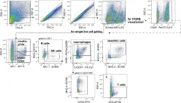
The liver is a complex organ that governs many types of metabolisms, including energy metabolism and other cellular processes. The liver also plays a crucial role in important functions in immunity, and the activity of liver tissue-associated immunity affects the outcome of many liver pathologies. A thorough characterization of the liver immune microenvironment may contribute to a better understanding of immune signaling, the mechanisms of specific immune responses, and even to improved predictions about therapy outcomes. In this paper, we present an optimized, simple, and rapid protocol to characterize the liver-associated immune cell milieu. We believe that the most suitable technique for obtaining a complex immune cell suspension and for removing contaminating blood cells is to perform mouse liver perfusion, using only phosphate buffer saline. Combining an enzymatic digestion and a mechanical dissociation of liver tissue, followed by cell purification, improves downstream applications. This combination is an essential prerequisite for immune cell determination and characterization. We then demonstrate a flow cytometry-based multiparametric immunophenotyping along with a gating strategy to detect and quantify liver endothelial cells, T cells (helper and cytotoxic), B cells, NK cells, NKT cells, neutrophils, monocytes (subsets included), dendritic cells (subsets included), macrophages and Kupffer cells.
-
Immunology and Microbiology
In Elife on 14 December 2022 by Pleuger, C., Ai, D., et al.
Fig.2.A

-
FC/FACS
-
Mus musculus (House mouse)
Collected and cropped from Elife by CiteAb, provided under a CC-BY license
Image 1 of 1