Bacille Calmette-Guérin (BCG) vaccination has off-target effects on disease risk for unrelated infections and immune responses to vaccines. This study aimed to determine the immunomodulatory effects of BCG vaccination on immune responses to vaccines against SARS-CoV-2.
Blood samples, from a subset of 275 SARS-CoV-2-naïve healthcare workers randomised to BCG vaccination (BCG group) or no BCG vaccination (Control group) in the BRACE trial, were collected before and 28 days after the primary course (two doses) of ChAdOx1-S (Oxford-AstraZeneca) or BNT162b2 (Pfizer-BioNTech) vaccination. SARS-CoV-2-specific antibodies were measured using ELISA and multiplex bead array, whole blood cytokine responses to γ-irradiated SARS-CoV-2 (iSARS) stimulation were measured by multiplex bead array, and SARS-CoV-2-specific T-cell responses were measured by activation-induced marker and intracellular cytokine staining assays.
After randomisation (mean 11 months) but prior to COVID-19 vaccination, the BCG group had lower cytokine responses to iSARS stimulation than the Control group. After two doses of ChAdOx1-S, differences in iSARS-induced cytokine responses between the BCG group and Control group were found for three cytokines (CTACK, TRAIL and VEGF). No differences were found between the groups after BNT162b2 vaccination. There were also no differences between the BCG and Control groups in COVID-19 vaccine-induced antigen-specific antibody responses, T-cell activation or T-cell cytokine production.
BCG vaccination induced a broad and persistent reduction in ex vivo cytokine responses to SARS-CoV-2. Following COVID-19 vaccination, this effect was abrogated, and BCG vaccination did not influence adaptive immune responses to COVID-19 vaccine antigens.
© 2025 The Author(s). Clinical & Translational Immunology published by John Wiley & Sons Australia, Ltd on behalf of Australian and New Zealand Society for Immunology, Inc.
Product Citations: 15
In Clinical Translational Immunology on 28 January 2025 by Messina, N. L., Germano, S., et al.
-
COVID-19
-
Immunology and Microbiology
In Molecular Therapy. Nucleic Acids on 10 September 2024 by Delehedde, C., Ciganek, I., et al.
mRNA applications have undergone unprecedented applications-from vaccination to cell therapy. Natural killer (NK) cells are recognized to have a significant potential in immunotherapy. NK-based cell therapy has drawn attention as allogenic graft with a minimal graft-versus-host risk leading to easier off-the-shelf production. NK cells can be engineered with either viral vectors or electroporation, involving high costs, risks, and toxicity, emphasizing the need for alternative way as mRNA technology. We successfully developed, screened, and optimized novel lipid-based platforms based on imidazole lipids. Formulations are produced by microfluidic mixing and exhibit a size of approximately 100 nm with a polydispersity index of less than 0.2. They are able to transfect NK-92 cells, KHYG-1 cells, and primary NK cells with high efficiency without cytotoxicity, while Lipofectamine Messenger Max and D-Lin-MC3 lipid nanoparticle-based formulations do not. Moreover, the translation of non-modified mRNA was higher and more stable in time compared with a modified one. Remarkably, the delivery of therapeutically relevant interleukin 2 mRNA resulted in extended viability together with preserved activation markers and cytotoxic ability of both NK cell lines and primary NK cells. Altogether, our platforms feature all prerequisites needed for the successful deployment of NK-based therapeutic strategies.
© 2024 The Authors.
-
Homo sapiens (Human)
-
Genetics
Preprint on BioRxiv : the Preprint Server for Biology on 28 March 2024 by Neuwirth, T., Malzl, D., et al.
Summary Regulatory T cells (T regs ) are a critical immune component guarding against excessive inflammatory responses. During chronic inflammation, T regs fail to control effector T cell responses. The causes of T reg dysfunction in these diseases are poorly characterized and therapies are aimed at blocking aberrant effector responses rather than rescuing T reg function. Here we utilized single-cell RNA sequencing data from patients suffering from chronic skin and colon inflammation to uncover SAT1 , the gene encoding spermidine/spermine N1-acetyltransferase (SSAT), as a novel marker and driver of skin-specific T reg dysfunction during T H 17-mediated inflammation. T regs expressing SAT1 exhibit a tissue-specific inflammation signature and show a proinflammatory effector-like profile. In CRISPRa on healthy human skin-derived T regs increased expression of SAT1 leads to a loss of suppressive function and a switch to a T H 17-like phenotype. This phenotype is induced by co-receptor expression on keratinocytes exposed to a T H 17 microenvironment. Finally, the potential therapeutic impact of targeting SSAT was demonstrated in a mouse model of skin inflammation by inhibiting SSAT pharmacologically, which rescued T reg number and function in the skin and systemically. Together, these data show that SAT1 expression has severe functional consequences on T regs and provides a novel target to treat chronic inflammatory skin disease.
-
FC/FACS
-
Immunology and Microbiology
In Comparative Medicine on 27 August 2023 by Christensen, P. K., Hansen, A. K., et al.
Immunodeficient mice engrafted with psoriatic human skin are widely used for the preclinical evaluation of new drug candidates. However, the T-cell activity, including the IL23/IL17 pathway, declines in the graft over time after engraftment, which likely affects the study data. Here, we investigated whether the T-cell activity could be sustained in xenografted psoriatic skin by local stimulation of T cells or systemic injection of autologous CD4 + T cells. We surgically transplanted human psoriatic skin from 5 untreated patients onto female NOG mice. Six days after surgery, mice received an intraperitoneal injection of autologous human CD4+ T cells, a subcutaneous injection under the grafts of a T-cell stimulation cocktail consisting of recombinant human IL2, human IL23, antihuman CD3, and antihuman CD28, or saline. Mice were euthanized 21 d after surgery and spleens and graft biopsies were collected for analysis. Human T cells were present in the grafts, and 60% of the grafts maintained the psoriatic phenotype. However, neither local T-cell stimulation nor systemic injection of autologous CD4+ T cells affected the protein levels of human IL17A, IL22, IFN γ, and TNF α in the grafts. In conclusion, NOG mice seem to accept psoriatic skin grafts, but the 2 approaches studied here did not affect human T-cell activity in the grafts. Therefore, NOG mice do not appear in this regard to be superior to other immunodeficient mice used for psoriasis xenografts.
-
Immunology and Microbiology
In Nature Immunology on 1 June 2023 by Zhang, W., Kedzierski, L., et al.
High-risk groups, including Indigenous people, are at risk of severe COVID-19. Here we found that Australian First Nations peoples elicit effective immune responses to COVID-19 BNT162b2 vaccination, including neutralizing antibodies, receptor-binding domain (RBD) antibodies, SARS-CoV-2 spike-specific B cells, and CD4+ and CD8+ T cells. In First Nations participants, RBD IgG antibody titers were correlated with body mass index and negatively correlated with age. Reduced RBD antibodies, spike-specific B cells and follicular helper T cells were found in vaccinated participants with chronic conditions (diabetes, renal disease) and were strongly associated with altered glycosylation of IgG and increased interleukin-18 levels in the plasma. These immune perturbations were also found in non-Indigenous people with comorbidities, indicating that they were related to comorbidities rather than ethnicity. However, our study is of a great importance to First Nations peoples who have disproportionate rates of chronic comorbidities and provides evidence of robust immune responses after COVID-19 vaccination in Indigenous people.
© 2023. The Author(s).
-
COVID-19
-
Immunology and Microbiology
In Nature on 1 April 2021 by Platten, M., Bunse, L., et al.
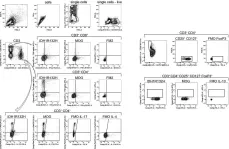
Fig.4.C

-
FC/FACS
-
Homo sapiens (Human)
Collected and cropped from Nature by CiteAb, provided under a CC-BY license
Image 1 of 1
