Premetastatic cancer cells often spread from the primary lesion through the lymphatic vasculature and, clinically, the presence or absence of lymph node metastases impacts treatment decisions. However, little is known about cancer progression via the lymphatic system or of the effect that the lymphatic environment has on cancer progression. This is due, in part, to the technical challenge of studying lymphatic vessels and collecting lymph fluid. Here we provide a step-by-step procedure to collect both lymph and tumor-draining lymph in mouse models of cancer metastasis. This protocol has been adapted from established methods of lymph collection and was developed specifically for the collection of lymph from tumors. The approach involves the use of mice bearing melanoma or breast cancer orthotopic tumors. After euthanasia, the cisterna chyli and the tumor are exposed and viewed using a stereo microscope. Then, a glass cannula connected to a 1 mL syringe is inserted directly into the cisterna chyli or the tumor-draining lymphatics for collection of pure lymph. These lymph samples can be used to analyze the lymph-derived cancer cells using highly sensitive multiomics approaches to investigate the impact of the lymph environment during cancer metastasis. The procedure requires 2 h per mouse to complete and is suitable for users with minimal expertise in small animal handling and use of microsurgical tools under a stereo microscope.
© 2025. Springer Nature Limited.
Product Citations: 29
Lymphatic collection and cell isolation from mouse models for multiomic profiling.
In Nature Protocols on 1 April 2025 by Sabatier, M., Solanki, A., et al.
In Nature Protocols on 1 March 2025 by Ma, L., Thapa, B. R., et al.
Durable and functional regeneration of the airway epithelium in vivo with transplanted stem cells has the potential to reconstitute healthy tissue in diseased airways, such as in cystic fibrosis or primary ciliary dyskinesia. Here, we present detailed protocols for the preparation and culture expansion of murine primary and induced pluripotent stem cell-derived airway basal stem cells (iBCs) and methods for their intra-airway transplantation into polidocanol-conditioned murine recipients to achieve durable in vivo airway regeneration. Reconstitution of the airway tissue resident epithelial stem cell compartment of immunocompetent mice with syngeneic donor cells leverages the extensive self-renewal and multipotent differentiation properties of basal stem cells (BCs) to durably generate a broad diversity of mature airway epithelial lineages in vivo. Engrafted donor-derived cells re-establish planar cell polarity as well as functional ciliary transport. By using this same approach, human primary BCs or iBCs transplanted into NOD-SCID gamma recipient mice similarly display engraftment and multilineage airway epithelial differentiation in vivo. The time to generate mouse or human iBCs takes ~60 d, which can be reduced to ~20 d if previously differentiated cells are thawed from cryopreserved iBC archives. The tracheal conditioning regimen and cell transplantation procedure is completed in 1 d. A competent graduate student or postdoctoral trainee should be able to perform the procedures listed in this protocol.
© 2024. Springer Nature Limited.
-
Stem Cells and Developmental Biology
In mBio on 11 December 2024 by Liu, H., Lin, J., et al.
The epidermal growth factor receptor (EGFR) has been identified as an epithelial cell receptor for Mucorales fungi and Candida albicans. Blocking EGFR with small molecule inhibitors reduces disease severity in mouse models of mucormycosis and oropharyngeal candidiasis. In contrast, cases of invasive aspergillosis have been reported in cancer patients who were treated with EGFR inhibitors, suggesting that EGFR signaling may play a protective role in the host defense against this infection. Here, we analyzed transcriptomic data from the lungs of mice with invasive aspergillosis and found evidence that Aspergillus fumigatus infection activates multiple genes that are predicted to function in the EGFR signaling pathway. We also found that A. fumigatus infection activates EGFR in both a human small-airway epithelial (HSAE) cell line and in the lungs of immunosuppressed mice. EGFR signaling in HSAE cells is required for maximal endocytosis of A. fumigatus and for fungal-induced proinflammatory cytokine and chemokine production. In a corticosteroid immunosuppressed mouse model of invasive pulmonary aspergillosis, inhibition of EGFR with gefitinib decreased whole-lung cytokine and chemokine levels and reduced accumulation of phagocytes in the lung, leading to a decrease in fungal killing, an increase in pulmonary fungal burden, and accelerated mortality. Thus, EGFR signaling is required for pulmonary epithelial cells to orchestrate the host innate immune defense against invasive aspergillosis in immunosuppressed hosts.IMPORTANCEWhen A. fumigatus infects the lungs, it invades epithelial cells that line the airways. During this process, the fungus interacts with epithelial cell receptors. This interaction stimulates epithelial cells to endocytose the fungus. It also induces these cells to secrete proinflammatory cytokines and chemokines that recruit phagocytes to the site of infection where they can kill the fungus. Here, we show that in small-airway epithelial cells, the EGFR acts as a sensor for A. fumigatus that triggers the production of chemokines in response to fungal infection. In corticosteroid-immunosuppressed mice, blocking EGFR with the kinase inhibitor gefitinib reduces chemokine production in the lungs. This leads to decreased accumulation of neutrophils and dendritic cells in the lungs, reduced A. fumigatus killing, and increased mortality. These results provide a potential explanation as to why some cancer patients who are treated with EGFR inhibitors develop invasive aspergillosis.
-
Mus musculus (House mouse)
-
Immunology and Microbiology
Preprint on BioRxiv : the Preprint Server for Biology on 10 September 2024 by Liu, H., Lin, J., et al.
The epidermal growth factor receptor (EGFR) has been identified as an epithelial cell receptor for Mucorales fungi and Candida albicans . Blocking EGFR with small molecule inhibitors reduces disease severity in mouse models of mucormycosis and oropharyngeal candidiasis. In contrast, cases of invasive aspergillosis have been reported in cancer patients who were treated with EGFR inhibitors, suggesting that EGFR signaling may play a protective role in the host defense against this infection. Here, we analyzed transcriptomic data from the lungs of mice with invasive aspergillosis and found evidence that Aspergillus fumigatus infection activates multiple genes that are predicted to function in the EGFR signaling pathway. We also found that A. fumigatus infection activates EGFR in both a human small airway epithelial (HSAE) cell line and in the lungs of immunosuppressed mice. EGFR signaling in HSAE cells is required for maximal endocytosis of A. fumigatus and for fungal-induced proinflammatory cytokine and chemokine production. In a corticosteroid immunosuppressed mouse model of invasive pulmonary aspergillosis, inhibition of EGFR with gefitinib decreased whole lung chemokine levels and reduced accumulation of phagocytes in the lung, leading to a decrease in fungal killing, an increase in pulmonary fungal burden, and accelerated mortality. Thus, EGFR signaling is required for pulmonary epithelial cells to orchestrate the host innate immune defense against invasive aspergillosis in immunosuppressed hosts. Importance When A. fumigatus infects the lungs, it invades epithelial cells that line the airways. During this process, the fungus interacts with epithelial cell receptors. This interaction stimulates epithelial cells to endocytose the fungus. It also induces these cells to secret proinflammatory cytokines and chemokines that recruit phagocytes to the site of infection where they can kill the fungus. Here, we show that in small airway epithelial cells, the epidermal growth factor receptor (EGFR) acts a sensor for A. fumigatus that triggers the production of chemokines in response to fungal infection. In corticosteroid-immunosuppressed mice, blocking EGFR with the kinase inhibitor, gefitinib reduces chemokine production in the lungs. This leads to decreased accumulation of neutrophils and dendritic cell in the lungs, reduced A. fumigatus killing, and increased mortality. These results provide a potential explanation as to why some cancer patients who are treated with EGFR inhibitors develop invasive aspergillosis.
-
Mus musculus (House mouse)
-
Immunology and Microbiology
Senescent fibroblasts in the tumor stroma rewire lung cancer metabolism and plasticity
Preprint on BioRxiv : the Preprint Server for Biology on 30 July 2024 by Lee, J. Y., Reyes, N., et al.
Summary Senescence has been demonstrated to either inhibit or promote tumorigenesis. Resolving this paradox requires spatial mapping and functional characterization of senescent cells in the native tumor niche. Here, we identified senescent p16 Ink4a + cancer-associated fibroblasts with a secretory phenotype that promotes fatty acid uptake and utilization by aggressive lung adenocarcinoma driven by Kras and p53 mutations. Furthermore, rewiring of lung cancer metabolism by p16 Ink4a + cancer- associated fibroblasts also altered tumor cell identity to a highly plastic/dedifferentiated state associated with progression in murine and human LUAD. Our ex vivo senolytic screening platform identified XL888, a HSP90 inhibitor, that cleared p16 Ink4a + cancer- associated fibroblasts in vivo . XL888 administration after establishment of advanced lung adenocarcinoma significantly reduced tumor burden concurrent with the loss of plastic tumor cells. Our study identified a druggable component of the tumor stroma that fulfills the metabolic requirement of tumor cells to acquire a more aggressive phenotype.
-
Mus musculus (House mouse)
-
Biochemistry and Molecular biology
-
Cancer Research
-
Cell Biology
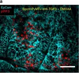
In Nat Commun on 11 September 2019 by Guerin, M. V., Regnier, F., et al.
Fig.6.A

-
IHC
-
Mus musculus (House mouse)
Collected and cropped from Nat Commun by CiteAb, provided under a CC-BY license
Image 1 of 1