Abstract Background. X-linked retinoschisis is a retinovitreal disorder primarily affecting males, starting in childhood. Over time, patients experience deterioration of vision due to the lack of retinoschisin-1 function. In clinical trials performing ocular gene delivery in those affected by this disorder, ocular inflammation was observed, which may have masked efficacy. A subsequent study focusing on analyzing the populations of peripheral blood mononuclear cells and cytokines in adults with this disease reported aberrant dendritic cell numbers and cytokine levels in peripheral blood. Adults with this disease may have an altered baseline immunity. However, children with X-linked retinoschisis were not included in that study, and whether the aberrant peripheral immunity in affected adults was a consequence of advanced eye pathology remained unclear. This study focuses on analyzing the populations of blood lymphocyte subsets in children aged 0 to 7 with X-linked retinoschisis and age-matched controls using flow cytometry. Results. The fractions of lymphocyte subsets that were CD16a + were significantly lower in children with X-linked retinoschisis. The fractions of lymphocyte subsets that were CD16a+/CD56+, namely natural killer cells, were also significantly lower. In children with X-linked retinoschisis, the fractions of CD3+/CD4 + T cells were higher, and the fractions of CD3 + CD8 + T cells were lower, despite having the same amounts of total CD3 + T cells within their lymphocyte populations. This resulted in a significantly greater CD4/CD8 ratio in children with X-linked retinoschisis compared to age-matched controls. Conclusions. Alterations were found in blood lymphocyte compositions of children with X-linked retinoschisis within both innate and adaptive immune axes. Some alterations including an elevation of CD4/CD8 ratio in X-linked retinoschisis mirror those previously found in adult patients with this disease. The fact that these abnormalities were present early in this disease indicates that retinoschisin-1 may play a role in regulating immunity in addition to retinal adhesion. The findings may have implications for future treatments such as ocular gene delivery.
Product Citations: 23
Immune landscape in children with X-linked retinoschisis
Preprint on Research Square on 17 April 2025 by Hsu, Y., Valle, G. D., et al.
-
Immunology and Microbiology
In Nature Communications on 10 March 2025 by Kgoadi, K., Bajpai, P., et al.
People living with HIV (PLWH) have an increased risk for developing tuberculosis after M. tuberculosis infection, despite anti-retroviral therapy (ART). To delineate the underlying mechanisms, we conducted single cell transcriptomics on bronchoalveolar lavage cells from PLWH on ART and HIV uninfected healthy controls infected with M. tuberculosis ex vivo. We identify an M1-like proinflammatory alveolar macrophage subset that sequentially acquires TNF signaling capacity in controls but not in PLWH. Cell-cell communication analyses reveal interactions between M1-like macrophages and effector memory T cells within TNF superfamily, chemokine, and costimulatory networks in the airways of controls. These interaction networks were lacking in PLWH infected with M. tuberculosis, where anti-inflammatory M2-like alveolar macrophages and T regulatory cells dominated along with dysregulated T cell signatures. Our data support a model in which impaired TNF-TNFR signaling, M2-like alveolar macrophages and aberrant macrophage-T cell crosstalk, lead to ineffective immunity to M. tuberculosis in PLWH on ART.
© 2025. The Author(s).
-
FC/FACS
-
Homo sapiens (Human)
-
Immunology and Microbiology
In Acta Pharmaceutica Sinica. B on 1 August 2024 by Niu, B., Tian, T., et al.
SMAD4 deficiency in colorectal cancer (CRC) is highly correlated with liver metastasis and high mortality, yet there are few effective precision therapies available. Here, we show that CCR1+-granulocytic myeloid-derived suppressor cells (G-MDSCs) are highly infiltrated in SMAD4-deficient CRC via CCL15/CCR1 and CCL9/CCR1 axis in clinical specimens and mouse models, respectively. The excessive TGF-β, secreted by tumor-infiltrated CCR1+-G-MDSCs, suppresses the immune response of cytotoxic T lymphocytes (CTLs), thus facilitating metastasis. Hereby, we develop engineered nanovesicles displaying CCR1 and TGFBR2 molecules (C/T-NVs) to chemotactically target the tumor driven by CCL9/CCR1 axis and trap TGF-β through TGF-β-TGFBR2 specific binding. Chemotactic C/T-NVs counteract CCR1+-G-MDSC infiltration through competitive responding CCL9/CCR1 axis. C/T-NVs-induced intratumoral TGF-β exhaustion alleviates the TGF-β-suppressed immune response of CTLs. Collectively, C/T-NVs attenuate liver metastasis of SMAD4-deficient CRC. In further exploration, high expression of programmed cell death ligand-1 (PD-L1) is observed in clinical specimens of SMAD4-deficient CRC. Combining C/T-NVs with anti-PD-L1 antibody (aPD-L1) induces tertiary lymphoid structure formation with sustained activation of CTLs, CXCL13+-CD4+ T, CXCR5+-CD20+ B cells, and enhanced secretion of cytotoxic cytokine interleukin-21 and IFN-γ around tumors, thus eradicating metastatic foci. Our strategy elicits pleiotropic antimetastatic immunity, paving the way for nanovesicle-mediated precision immunotherapy in SMAD4-deficient CRC.
© 2024 The Authors.
-
FC/FACS
-
Cancer Research
In Cell Stem Cell on 1 August 2024 by Jakobsen, N. A., Turkalj, S., et al.
Clonal hematopoiesis (CH) arises when hematopoietic stem cells (HSCs) acquire mutations, most frequently in the DNMT3A and TET2 genes, conferring a competitive advantage through mechanisms that remain unclear. To gain insight into how CH mutations enable gradual clonal expansion, we used single-cell multi-omics with high-fidelity genotyping on human CH bone marrow (BM) samples. Most of the selective advantage of mutant cells occurs within HSCs. DNMT3A- and TET2-mutant clones expand further in early progenitors, while TET2 mutations accelerate myeloid maturation in a dose-dependent manner. Unexpectedly, both mutant and non-mutant HSCs from CH samples are enriched for inflammatory and aging transcriptomic signatures, compared with HSCs from non-CH samples, revealing a non-cell-autonomous effect. However, DNMT3A- and TET2-mutant HSCs have an attenuated inflammatory response relative to wild-type HSCs within the same sample. Our data support a model whereby CH clones are gradually selected because they are resistant to the deleterious impact of inflammation and aging.
Copyright © 2024 The Author(s). Published by Elsevier Inc. All rights reserved.
-
Immunology and Microbiology
-
Stem Cells and Developmental Biology
In Cell Reports on 23 July 2024 by Pelletier, A. N., Sánchez, G. P., et al.
Protective immunity to dengue virus (DENV) requires antibody response to all four serotypes. Systems vaccinology identifies a multi-OMICs pre-vaccination signature and mechanisms predictive of broad antibody responses after immunization with a tetravalent live attenuated DENV vaccine candidate (Butantan-DV/TV003). Anti-inflammatory pathways, including TGF-β signaling expressed by CD68low monocytes, and the metabolites phosphatidylcholine (PC) and phosphatidylethanolamine (PE) positively correlate with broadly neutralizing antibody responses against DENV. In contrast, expression of pro-inflammatory pathways and cytokines (IFN and IL-1) in CD68hi monocytes and primary and secondary bile acids negatively correlates with broad DENV-specific antibody responses. Induction of TGF-β and IFNs is done respectively by PC/PE and bile acids in CD68low and CD68hi monocytes. The inhibition of viral sensing by PC/PE-induced TGF-β is confirmed in vitro. Our studies show that the balance between metabolites and the pro- or anti-inflammatory state of innate immune cells drives broad and protective B cell response to a live attenuated dengue vaccine.
Copyright © 2024 The Authors. Published by Elsevier Inc. All rights reserved.
-
FC/FACS
-
Biochemistry and Molecular biology
-
Cell Biology
-
Immunology and Microbiology
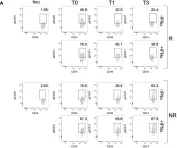
In Front Immunol on 13 August 2022 by Tucci, G., Garufi, C., et al.
Fig.3.A

-
FC/FACS
-
Homo sapiens (Human)
Collected and cropped from Front Immunol by CiteAb, provided under a CC-BY license
Image 1 of 1