Despite advances in immunotherapy, metastatic melanoma remains a considerable therapeutic challenge due to the complexity of the tumor microenvironment. Intratumoral type I interferon (IFN-I) has long been associated with improved clinical outcomes. However, several IFN-I subtypes can also paradoxically promote tumor growth in some contexts. We investigated this further by engineering murine B16 melanoma cells to overexpress various IFN-I subtypes, where a spectrum of outcomes was observed. Characterization of these tumors by RNA sequencing revealed a tumor immune phenotype, where potent IFN-I signaling concomitant with diminished type 2 inflammation failed to confer durable tumor control. T cell-mediated rejection of these tumors was restored by introducing interleukin-4 (IL-4) into the tumor microenvironment, either through ectopic expression or in a preclinical adoptive T cell therapy model. Collectively, our findings highlight the IFN-I/IL-4 axis in promoting antitumor immunity, which could be harnessed to target and stratify solid tumors that are nonresponsive to frontline therapies.
Product Citations: 30
Interleukin-4 modulates type I interferon to augment antitumor immunity.
In Science Advances on 16 May 2025 by Newnes, H. V., Armitage, J. D., et al.
-
Immunology and Microbiology
In The Journal of Pathology on 1 January 2025 by Milan, M., Maiullari, F., et al.
Duchenne muscular dystrophy (DMD) is caused by the absence of the full form of the dystrophin protein, which is essential for maintaining the structural integrity of muscle cells, including those in the heart and respiratory system. Despite progress in understanding the molecular mechanisms associated with DMD, myocardial insufficiency persists as the primary cause of mortality, and existing therapeutic strategies remain limited. This study investigates the hypothesis that a dysregulation of the biological communication between infiltrating macrophages (MPs) and neurocardiac junctions exists in dystrophic cardiac tissue. In a mouse model of DMD (mdx), this phenomenon is influenced by the over-release of chondroitin sulfate proteoglycan-4 (CSPG4), a key inhibitor of nerve sprouting and a modulator of the neural function, by MPs infiltrating the cardiac tissue and associated with dilated cardiomyopathy, a hallmark of DMD. Givinostat, the histone deacetylase inhibitor under current development as a clinical treatment for DMD, is effective at both restoring a physiological microenvironment at the neuro-cardiac junction and cardiac function in mdx mice in addition to a reduction in cardiac fibrosis, MP-mediated inflammation, and tissue CSPG4 content. This study provides novel insight into the pathophysiology of DMD in the heart, identifying potential new biological targets. © 2024 The Author(s). The Journal of Pathology published by John Wiley & Sons Ltd on behalf of The Pathological Society of Great Britain and Ireland.
© 2024 The Author(s). The Journal of Pathology published by John Wiley & Sons Ltd on behalf of The Pathological Society of Great Britain and Ireland.
-
Mus musculus (House mouse)
-
Cardiovascular biology
-
Pathology
In International Journal of Molecular Sciences on 19 December 2024 by Akhter, N., Contreras, J., et al.
Traumatic optic neuropathy (TON) has been regarded a vision-threatening condition caused by either ocular or blunt/penetrating head trauma, which is characterized by direct or indirect TON. Injury happens during sports, vehicle accidents and mainly in military war and combat exposure. Earlier, we have demonstrated that remote ischemic post-conditioning (RIC) therapy is protective in TON, and here we report that AMPKα1 activation is crucial. AMPKα1 is the catalytic subunit of the heterotrimeric enzyme AMPK, the master regulator of cellular energetics and metabolism. The α1 isoform predominates in immune cells including macrophages (Mφs). Myeloid-specific AMPKα1 KO mice were generated by crossing AMPKα1Flox/Flox and LysMcre to carry out the study. We induced TON in mice by using a controlled impact system. Mice (mixed sex) were randomized in six experimental groups for Sham (mock); Sham (RIC); AMPKα1F/F (TON); AMPKα1F/F (TON+RIC); AMPKα1F/F LysMCre (TON); AMPKα1F/F LysMCre (TON+RIC). RIC therapy was given every day (5-7 days following TON). Data were generated by using Western blotting (pAMPKα1, ICAM1, Brn3 and GAP43), immunofluorescence (pAMPKα1, cd11b, TMEM119 and ICAM1), flow cytometry (CD11b, F4/80, CD68, CD206, IL-10 and LY6G), ELISA (TNF-α and IL-10) and transmission electron microscopy (TEM, for demyelination and axonal degeneration), and retinal oxygenation was measured by a Unisense sensor system. First, we observed retinal morphology with funduscopic images and found TON has vascular inflammation. H&E staining data suggested that TON increased retinal inflammation and RIC attenuates retinal ganglion cell death. Immunofluorescence and Western blot data showed increased microglial activation and decreased retinal ganglion cell (RGCs) marker Brn3 and axonal regeneration marker GAP43 expression in the TON [AMPKα1F/F] vs. Sham group, but TON+RIC [AMPKα1F/F] attenuated the expression level of these markers. Interestingly, higher microglia activation was observed in the myeloid AMPKα1F/F KO group following TON, and RIC therapy did not attenuate microglial expression. Flow cytometry, ELISA and retinal tissue oxygen data revealed that RIC therapy significantly reduced the pro-inflammatory signaling markers, increased anti-inflammatory macrophage polarization and improved oxygen level in the TON+RIC [AMPKα1F/F] group; however, RIC therapy did not reduce inflammatory signaling activation in the myeloid AMPKα1 KO mice. The transmission electron microscopy (TEM) data of the optic nerve showed increased demyelination and axonal degeneration in the TON [AMPKα1F/F] group, and RIC improved the myelination process in TON [AMPKα1F/F], but RIC had no significant effect in the AMPKα1 KO mice. The myeloid AMPKα1c deletion attenuated RIC induced anti-inflammatory macrophage polarization, and that suggests a molecular link between RIC and immune activation. Overall, these data suggest that RIC therapy provided protection against inflammation and neurodegeneration via myeloid AMPKα1 activation, but the deletion of myeloid AMPKα1 is not protective in TON. Further investigation of RIC and AMPKα1 signaling is warranted in TON.
-
Mus musculus (House mouse)
-
Immunology and Microbiology
In The Journal of Immunology on 1 December 2024 by Garcia-Flores, V., Liu, Z., et al.
Preterm birth (PTB), often preceded by preterm labor, is a major cause of neonatal morbidity and mortality worldwide. Most PTB cases involve intra-amniotic inflammation without detectable microorganisms, termed in utero sterile inflammation, for which there is no established treatment. In this study, we propose homeostatic macrophages to prevent PTB and adverse neonatal outcomes caused by in utero sterile inflammation. Single-cell atlases of the maternal-fetal interface revealed that homeostatic maternal macrophages are reduced with human labor. M2 macrophage treatment prevented PTB and reduced adverse neonatal outcomes in mice with in utero sterile inflammation. Specifically, M2 macrophages halted premature labor by suppressing inflammatory responses in the amniotic cavity, including inflammasome activation, and mitigated placental and offspring lung inflammation. Moreover, M2 macrophages boosted gut inflammation in neonates and improved their ability to fight systemic bacterial infections. Our findings show that M2 macrophages are a promising strategy to mitigate PTB and improve neonatal outcomes resulting from in utero sterile inflammation.
Copyright © 2024 by The American Association of Immunologists, Inc.
-
Immunology and Microbiology
In Cancer Immunology Research on 3 September 2024 by Franzolin, G., Brundu, S., et al.
Semaphorin-plexin signaling plays a major role in the tumor microenvironment (TME). In particular, Semaphorin 4D (SEMA4D) has been shown to promote tumor growth and metastasis; however, the role of its high-affinity receptor Plexin-B1 (PLXNB1), which is expressed in the TME, is poorly understood. In this study, we directly targeted PLXNB1 in the TME of triple-negative murine breast carcinoma to elucidate its relevance in cancer progression. We found that primary tumor growth and metastatic dissemination were strongly reduced in PLXNB1-deficient mice, which showed longer survival. PLXNB1 loss in the TME induced a switch in the polarization of tumor-associated macrophages (TAM) toward a pro-inflammatory M1 phenotype and enhanced the infiltration of CD8+ T lymphocytes both in primary tumors and in distant metastases. Moreover, PLXNB1 deficiency promoted a shift in the Th1/Th2 balance of the T-cell population and an antitumor gene signature, with the upregulation of Icos, Perforin-1, Stat3, and Ccl5 in tumor-infiltrating lymphocytes (TILs). We thus tested the translational relevance of TME reprogramming driven by PLXNB1 inactivation for responsiveness to immunotherapy. Indeed, in the absence of PLXNB1, the efficacy of anti-PD-1 blockade was strongly enhanced, efficiently reducing tumor growth and distant metastasis. Consistent with this, pharmacological PLXNB1 blockade by systemic treatment with a specific inhibitor significantly hampered breast cancer growth and enhanced the antitumor activity of the anti-PD-1 treatment in a preclinical model. Altogether, these data indicate that PLXNB1 signaling controls the antitumor immune response in the TME and highlight this receptor as a promising immune therapeutic target for metastatic breast cancers.
©2024 The Authors; Published by the American Association for Cancer Research.
-
Cancer Research
-
Immunology and Microbiology
In J Clin Invest on 1 November 2022 by de Souza, C. O., Paschoal, V. A., et al.
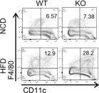
Fig.3.C

-
FC/FACS
-
Mus musculus (House mouse)
Collected and cropped from J Clin Invest by CiteAb, provided under a CC-BY license
Image 1 of 1