
Rho GTPases are key regulators of cell motility and membrane trafficking, influencing critical processes such as epithelial-mesenchymal transition (EMT). Among them, the small GTPase RhoB plays a pivotal role, but the mechanisms underlying its regulation remain largely unclear. We have previously identified the Rho guanine nucleotide exchange factor (RhoGEF) Solo (ARHGEF40) as a regulator of endosomal RhoB in epithelial cells. Here, we find that Solo is upregulated in breast cancer cells with high EMT scores and promotes cell motility through its RhoGEF activity. Solo's ability to enhance migration is further regulated by phosphorylation at tyrosine 242, mediated by the proto-oncogene Src. By combining high-resolution imaging with photoconversion assays, we further demonstrate that Solo regulates Src trafficking dynamics, localization, and consequently signaling at focal adhesions. Together, our data identify Solo as a novel feedback regulator of Src and a key driver of the motility of breast cancer cells with mesenchymal characteristics.
© 2025 The Author(s).
Product Citations: 113
In IScience on 20 June 2025 by Meyer, F., Lungu, C., et al.
In Molecular Omics on 12 May 2025 by Pascual-Vargas, P., Arias-Garcia, M., et al.
YAP and TAZ are transcriptional co-activators that are inhibited by sequestration in the cytoplasm. Cellular signalling pathways integrate soluble, mechanical (cytoskeleton, adhesion), and geometric (cell size, morphology) cues to regulate the translocation of YAP/TAZ to the nucleus. In triple-negative breast cancer (TNBC) cells, both signalling and morphogenesis are frequently rewired, leading to increased YAP/TAZ translocation, which drives proliferation, invasion, and drug resistance. However, whether this increased YAP/TAZ translocation is due to alterations in upstream signalling events or changes in cell morphology remains unclear. To gain insight into YAP/TAZ regulation in TNBC cells, we performed multiplexed quantitative genetic screens for YAP/TAZ localisation and cell shape, enabling us to determine whether changes in YAP/TAZ localisation following gene knockdown could be explained by alterations in cell morphology. These screens revealed that the focal adhesion (FA)-associated RhoGEF DOCK5 is essential for YAP/TAZ nuclear localisation in TNBC cells. DOCK5-defective cells exhibit defects in FA morphogenesis and fail to generate a stable, polarised leading edge, which we propose contributes to impaired YAP/TAZ translocation. Mechanistically, we implicate DOCK5's ability to act as a RacGEF and as a scaffold for NCK/AKT as key to its role in FA morphogenesis. Importantly, DOCK5 is essential for promoting the resistance of LM2 cells to the clinically used MEK inhibitor Binimetinib. Taken together, our findings suggest that DOCK5's role in TNBC cell shape determination drives YAP/TAZ upregulation and drug resistance.
Changes in nuclear and actin mechanics from G1 to G2 affect nuclear integrity
Preprint on BioRxiv : the Preprint Server for Biology on 16 April 2025 by Bunner, S., Huang, K., et al.
The structural integrity of the nucleus is dependent on nuclear mechanical elements of chromatin and lamins to resist antagonistic actin cytoskeleton forces. Imbalance results in nuclear blebbing, rupture, and cellular dysfunction found in many human diseases. We used Fluorescent Ubiquitin Cell Cycle Indicator (FUCCI) cells to determine how cell cycle changes affect the nucleus and actin force balance. While nuclear blebs are present equally throughout interphase, nuclear blebs form predominantly in G1 and then persist into G2 due to increased actin-based nuclear confinement and focal adhesion density in G1 vs. G2 cells. Upon artificial confinement, G2 nuclei ruptured more than G1 nuclei. Single nucleus micromanipulation force measurements confirmed that G1 nuclei are stronger than G2 nuclei in both the chromatin-based and lamin-based nuclear stiffness regimes. Decreased nuclear stiffness can be explained by loss of peripheral H3K9me3 from G1 to G2, recapitulated by H3K9me3 inhibition via Chaetocin. Cell cycle-based changes in nuclear and actin mechanics impact nuclear integrity and shape.
-
ICC-IF
-
Cell Biology
In ACS Applied Materials Interfaces on 22 January 2025 by Xiao, J., Ang, J. W., et al.
Focal adhesions (FAs) are force-bearing multiprotein complexes, whose nanoscale organization and signaling are essential for cell growth and differentiation. However, the specific organization of FA components to exert spatiotemporal activation of FA proteins for force sensing and transduction remains unclear. In this study, we unveil the intricacies of FA protein nanoarchitecture and that its dynamics are coordinated by a molecular scaffold protein, BNIP-2, to initiate downstream signal transduction for cardiomyoblast differentiation. Within the FAs, BNIP-2 regulates the nano-organization of focal adhesion kinase (FAK), and the dynamics of FAK, paxillin, and vinculin. Depletion of BNIP-2 resulted in altered focal adhesion numbers and sizes per cell, reduced traction force, and decreased FA sensitivity for mechanosensing. At the molecular level, the loss of BNIP-2 disrupted the FAK-paxillin signaling axis, where FAK inhibition reproduces the effects of BNIP-2 loss by impairing the phosphorylation of both FAK and paxillin. Mechanistically, BNIP-2 preferentially binds to constitutively active FAK and acts as a molecular scaffold to mediate interactions between FAK and paxillin and between paxillin and vinculin. We have validated BNIP-2's role in the FAK-paxillin signaling axis in human embryonic stem cells (hESC). Furthermore, we showed that depletion of BNIP-2 resulted in changes in signature gene targets at the cardiac progenitor stage of differentiation. In summary, we showed that the intricate interplay of FA nanoarchitecture and dynamics, governed by BNIP-2, is crucial for force transduction and biochemical signaling in driving cardiomyoblast differentiation.
A comparative analysis of paxillin and Hic-5 proximity interactomes.
In Cytoskeleton (Hoboken, N.J.) on 1 January 2025 by Brock, K., Alpha, K. M., et al.
Focal adhesions serve as structural and signaling hubs, facilitating bidirectional communication at the cell-extracellular matrix interface. Paxillin and the related Hic-5 (TGFβ1i1) are adaptor/scaffold proteins that recruit numerous structural and regulatory proteins to focal adhesions, where they perform both overlapping and discrete functions. In this study, paxillin and Hic-5 were expressed in U2OS osteosarcoma cells as biotin ligase (BioID2) fusion proteins and used as bait proteins for proximity-dependent biotinylation in order to directly compare their respective interactomes. The fusion proteins localized to both focal adhesions and the centrosome, resulting in biotinylation of components of each of these structures. Biotinylated proteins were purified and analyzed by mass spectrometry. The list of proximity interactors for paxillin and Hic-5 comprised numerous shared core focal adhesion proteins that likely contribute to their similar functions in cell adhesion and migration, as well as proteins unique to paxillin and Hic-5 that have been previously localized to focal adhesions, the centrosome, or the nucleus. Western blotting confirmed biotinylation and enrichment of FAK and vinculin, known interactors of Hic-5 and paxillin, as well as several potentially unique proximity interactors of Hic-5 and paxillin, including septin 7 and ponsin, respectively. Further investigation into the functional relationship between the unique interactors and Hic-5 or paxillin may yield novel insights into their distinct roles in cell migration.
© 2024 Wiley Periodicals LLC.
-
Homo sapiens (Human)
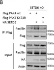
In Sci Rep on 13 October 2020 by Vershinin, Z., Feldman, M., et al.
Fig.5.B

-
WB
-
Homo sapiens (Human)
Collected and cropped from Sci Rep by CiteAb, provided under a CC-BY license
Image 1 of 11
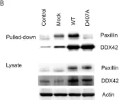
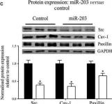
In Anim Cells Syst (Seoul) on 1 February 2019 by Sohn, S. O. & Chay, K. O.
Fig.2.B

-
WB
-
Collected and cropped from Anim Cells Syst (Seoul) by CiteAb, provided under a CC-BY license
Image 1 of 11
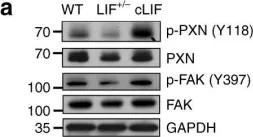
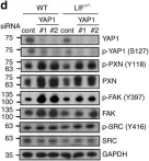
In Nat Commun on 30 November 2018 by Liu, S. C., Hsu, T., et al.
Fig.5.A

-
WB
-
Homo sapiens (Human)
Collected and cropped from Nat Commun by CiteAb, provided under a CC-BY license
Image 1 of 11
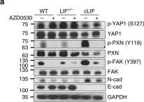
In Nat Commun on 30 November 2018 by Liu, S. C., Hsu, T., et al.
Fig.7.A

-
WB
-
Homo sapiens (Human)
Collected and cropped from Nat Commun by CiteAb, provided under a CC-BY license
Image 1 of 11
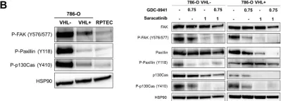
In Nat Commun on 30 November 2018 by Liu, S. C., Hsu, T., et al.
Fig.6.D

-
WB
-
Homo sapiens (Human)
Collected and cropped from Nat Commun by CiteAb, provided under a CC-BY license
Image 1 of 11
In Oncotarget on 10 July 2018 by Roelants, C., Giacosa, S., et al.
Fig.2.B

-
WB
-
Collected and cropped from Oncotarget by CiteAb, provided under a CC-BY license
Image 1 of 11
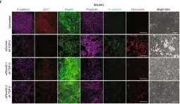
In Nat Commun on 15 November 2017 by Sahu, S. K., Tiwari, N., et al.
Fig.1.F

-
IF
-
Homo sapiens (Human)
Collected and cropped from Nat Commun by CiteAb, provided under a CC-BY license
Image 1 of 11
In J Cell Mol Med on 1 January 2017 by Nicholson, C. J., Seta, F., et al.
Fig.3.C

-
WB
-
Collected and cropped from J Cell Mol Med by CiteAb, provided under a CC-BY license
Image 1 of 11
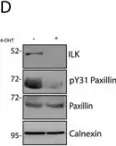
In J Cell Physiol on 1 November 2016 by Rooney, N., Wang, P., et al.
Fig.5.D

-
WB
-
Collected and cropped from J Cell Physiol by CiteAb, provided under a CC-BY license
Image 1 of 11
In J Cell Physiol on 1 November 2016 by Rooney, N., Wang, P., et al.
Fig.5.C

-
WB
-
Collected and cropped from J Cell Physiol by CiteAb, provided under a CC-BY license
Image 1 of 11
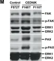
In PLoS One on 23 March 2010 by Rapaport, D., Lugassy, Y., et al.
Fig.7.M

-
WB
-
Homo sapiens (Human)
Collected and cropped from PLoS One by CiteAb, provided under a CC-BY license
Image 1 of 11