
Despite recent therapeutic advances, combating cancer resistance remains a formidable challenge. The 78-kilodalton glucose-regulated protein (GRP78), a key stress-inducible endoplasmic reticulum (ER) chaperone, plays a crucial role in both cancer cell survival and stress adaptation. GRP78 is also upregulated during SARS-CoV-2 infection and acts as a critical host factor. Recently, we discovered cardiac glycosides (CGs) as novel suppressors of GRP78 stress induction through a high-throughput screen of clinically relevant compound libraries. This study aims to test the possibility that agents capable of blocking stress induction of GRP78 could dually suppress cancer and COVID-19.
Here we report that oleandrin (OLN), is the most potent among the CGs in inhibiting acute stress induction of total GRP78, which also results in reduced cell surface and nuclear forms of GRP78 in stressed cells. The inhibition of stress induction of GRP78 is at the post-transcriptional level, independent of protein degradation and autophagy and may involve translational control as OLN blocks stress-induced loading of ribosomes onto GRP78 mRNAs. Moreover, the human Na+/K+-ATPase α3 isoform is critical for OLN suppression of GRP78 stress induction. OLN, in nanomolar range, enhances apoptosis, sensitizes colorectal cancer cells to chemotherapeutic agents, and reduces the viability of patient-derived colon cancer organoids. Likewise, OLN, suppresses GRP78 expression and impedes tumor growth in an orthotopic breast cancer xenograft model. Furthermore, OLN blocks infection by SARS-CoV-2 and its variants and enhances existing anti-viral therapies. Notably, GRP78 overexpression mitigates OLN-mediated cancer cell apoptotic onset and suppression of virus release.
Our findings validate GRP78 as a target of OLN anti-cancer and anti-viral activities. These proof-of-principle studies support further investigation of OLN as a readily accessible compound to dually combat cancer and COVID-19.
© 2024. The Author(s).
Product Citations: 41
In Cell Bioscience on 6 September 2024 by Ha, D. P., Shin, W. J., et al.
-
WB
-
Cancer Research
-
Cardiovascular biology
-
Cell Biology
-
COVID-19
In Matrix Biology : Journal of the International Society for Matrix Biology on 1 May 2024 by Long, A. M., Kwon, J. M., et al.
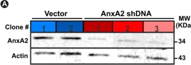
Extracellular matrix (ECM) pathologic remodeling underlies many disorders, including muscular dystrophy. Tissue decellularization removes cellular components while leaving behind ECM components. We generated "on-slide" decellularized tissue slices from genetically distinct dystrophic mouse models. The ECM of dystrophin- and sarcoglycan-deficient muscles had marked thrombospondin 4 deposition, while dysferlin-deficient muscle had excess decorin. Annexins A2 and A6 were present on all dystrophic decellularized ECMs, but annexin matrix deposition was excessive in dysferlin-deficient muscular dystrophy. Muscle-directed viral expression of annexin A6 resulted in annexin A6 in the ECM. C2C12 myoblasts seeded onto decellularized matrices displayed differential myoblast mobility and fusion. Dystrophin-deficient decellularized matrices inhibited myoblast mobility, while dysferlin-deficient decellularized matrices enhanced myoblast movement and differentiation. Myoblasts treated with recombinant annexin A6 increased mobility and fusion like that seen on dysferlin-deficient decellularized matrix and demonstrated upregulation of ECM and muscle cell differentiation genes. These findings demonstrate specific fibrotic signatures elicit effects on myoblast activity.
Copyright © 2024 The Authors. Published by Elsevier B.V. All rights reserved.
-
Mus musculus (House mouse)
In Journal of Extracellular Biology on 1 October 2023 by Hernandez, B. J., Skiba, N. P., et al.
The retinal pigmented epithelium (RPE) constitutes the outer blood-retinal barrier, enables photoreceptor function of the eye, and is constantly exposed to oxidative stress. As such, dysfunction of the RPE underlies pathology leading to development of age-related macular degeneration (AMD), the leading cause of vision loss among the elderly in industrialized nations. A major responsibility of the RPE is to process photoreceptor outer segments, which relies on the proper functioning of its endocytic pathways and endosomal trafficking. Exosomes and other extracellular vesicles (EVs) from RPE are an essential part of these pathways and may be early indicators of cellular stress. To test the role of small EVs (sEVs) including exosomes, that may underlie the early stages of AMD, we used a polarized primary RPE cell culture model under chronic subtoxic oxidative stress. Unbiased proteomic analyses of highly purified basolateral sEVs from oxidatively stressed RPE cultures revealed changes in proteins involved in epithelial barrier integrity. There were also significant changes in proteins accumulating in the basal-side sub-RPE extracellular matrix during oxidative stress, that could be prevented with an inhibitor of sEV release. Thus, chronic subtoxic oxidative stress in primary RPE cultures induces changes in sEV content, including basal-side specific desmosome and hemidesmosome shedding via sEVs. These findings provide novel biomarkers of early cellular dysfunction and opportunity for therapeutic intervention in age-related retinal diseases (e.g., AMD).
-
Cardiovascular biology
Preprint on BioRxiv : the Preprint Server for Biology on 13 June 2023 by Hernandez, B. J., Skiba, N. P., et al.
The retinal pigmented epithelium (RPE) constitutes the outer blood-retinal barrier, enables photoreceptor function of the eye, and is constantly exposed to oxidative stress. As such, dysfunction of the RPE underlies pathology leading to development of age-related macular degeneration (AMD), the leading cause of vision loss among the elderly in industrialized nations. A major responsibility of the RPE is to process photoreceptor outer segments, which relies on the proper functioning of its endocytic pathways and endosomal trafficking. Exosomes and other extracellular vesicles from RPE are an essential part of these pathways and may be early indicators of cellular stress. To test the role of exosomes that may underlie the early stages of AMD, we used a polarized primary RPE cell culture model under chronic subtoxic oxidative stress. Unbiased proteomic analyses of highly purified basolateral exosomes from oxidatively stressed RPE cultures revealed changes in proteins involved in epithelial barrier integrity. There were also significant changes in proteins accumulating in the basal-side sub-RPE extracellular matrix during oxidative stress, that could be prevented with an inhibitor of exosome release. Thus, chronic subtoxic oxidative stress in primary RPE cultures induces changes in exosome content, including basal-side specific desmosome and hemidesmosome shedding via exosomes. These findings provide novel biomarkers of early cellular dysfunction and opportunity for therapeutic intervention in age-related retinal diseases, (e.g., AMD) and broadly from blood-CNS barriers in other neurodegenerative diseases.
-
Cardiovascular biology
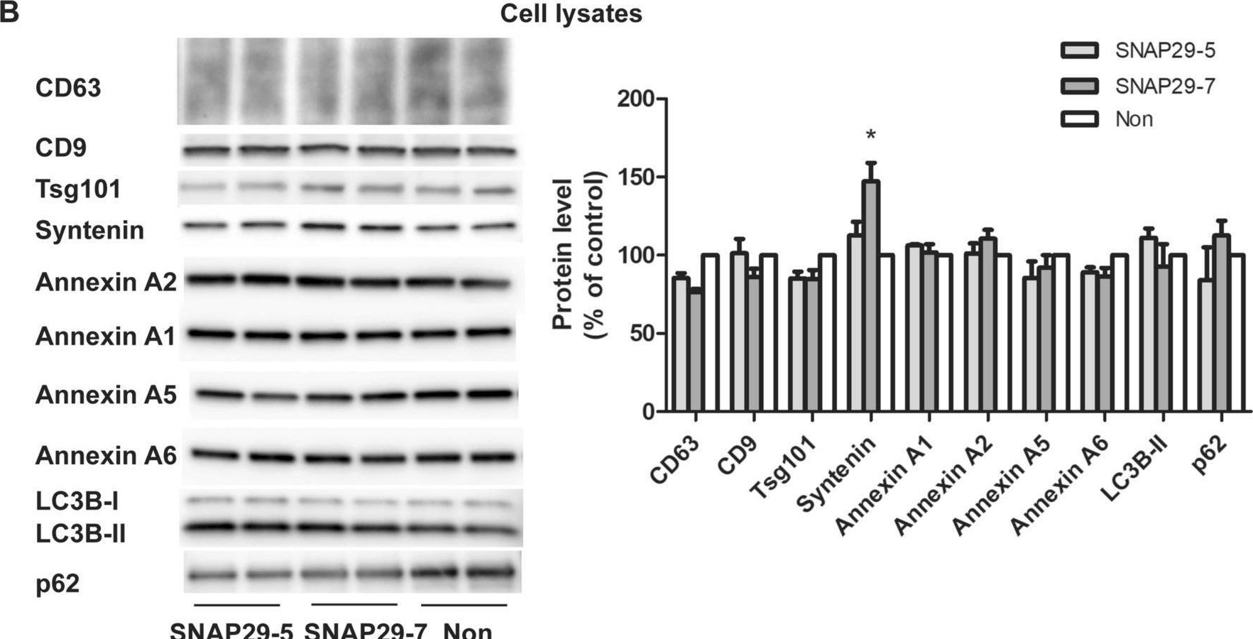
siRNA screening reveals that SNAP29 contributes to exosome release.
In Cellular and Molecular Life Sciences : CMLS on 7 June 2023 by Hessvik, N. P., Sagini, K., et al.
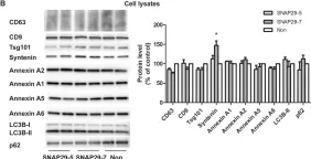
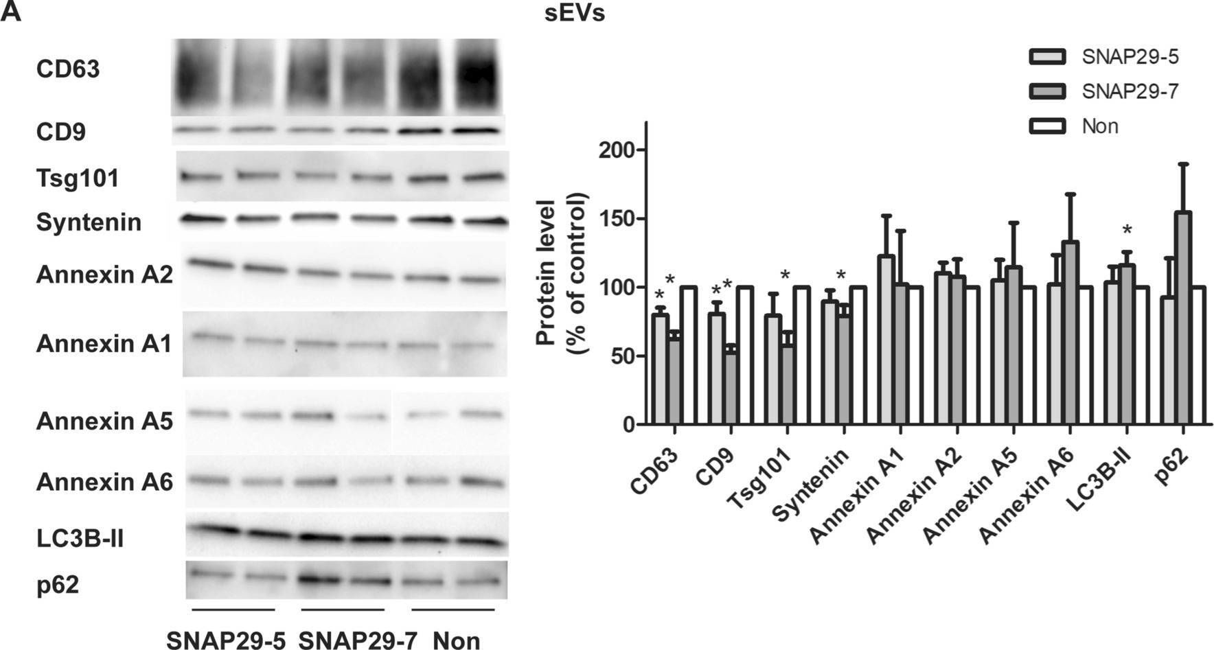
Cells release extracellular vesicles (EVs) of different sizes. Small EVs (< 200 nm) can originate from the fusion of multivesicular bodies with the plasma membrane, i.e. exosomes, and from budding of the plasma membrane, i.e. small ectosomes. To investigate the molecular machinery required for the release of small EVs, we developed a sensitive assay based on incorporation of radioactive cholesterol in EV membranes and used it in a siRNA screening. The screening showed that depletion of several SNARE proteins affected the release of small EVs. We focused on SNAP29, VAMP8, syntaxin 2, syntaxin 3 and syntaxin 18, the depletion of which reduced the release of small EVs. Importantly, this result was verified using gold standard techniques. SNAP29 depletion resulted in the largest effect and was further investigated. Immunoblotting analysis of small EVs showed that the release of several proteins considered to be associated with exosomes like syntenin, CD63 and Tsg101 was reduced, while the level of several proteins that have been shown to be released in ectosomes (annexins) or by secretory autophagy (LC3B and p62) was not affected by SNAP29 depletion. Moreover, these proteins appeared in different fractions when the EV samples were further separated by a density gradient. These results suggest that SNAP29 depletion mainly affects the secretion of exosomes. To investigate how SNAP29 affects exosome release, we used microscopy to study the distribution of MBVs using CD63 labelling and CD63-pHluorin to detect fusion events of MVBs with the plasma membrane. SNAP29 depletion caused a redistribution of CD63-labelled compartments but did not change the number of fusion events. Further experiments are therefore needed to fully understand the function of SNAP29. To conclude, we have developed a novel screening assay that has allowed us to identify several SNAREs involved in the release of small EVs.
© 2023. The Author(s).
-
WB
-
Biochemistry and Molecular biology
-
Genetics
In Cell Mol Life Sci on 7 June 2023 by Hessvik, N. P., Sagini, K., et al.
Fig.5.A
-
WB
-
Collected and cropped from Cell Mol Life Sci by CiteAb, provided under a CC-BY license
Image 1 of 4
In Cell Mol Life Sci on 7 June 2023 by Hessvik, N. P., Sagini, K., et al.
Fig.5.B
-
WB
-
Collected and cropped from Cell Mol Life Sci by CiteAb, provided under a CC-BY license
Image 1 of 4
In Cells on 19 August 2020 by Bittel, D. C., Chandra, G., et al.
Fig.2.A

-
WB
-
Collected and cropped from Cells by CiteAb, provided under a CC-BY license
Image 1 of 4
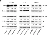
In PLoS One on 24 January 2018 by Gomes, S. E., Pereira, D. M., et al.
Fig.3.C

-
WB
-
Collected and cropped from PLoS One by CiteAb, provided under a CC-BY license
Image 1 of 4