Microglia, the resident immune cells of the central nervous system, play a pivotal role in maintaining homeostasis, responding to injury, and modulating neuroinflammation. However, the limitations of rodent models in accurately representing human microglia have posed significant challenges in the study of retinal diseases.
PLX5622 was used to eliminate endogenous microglia in mice through oral and intraperitoneal administration, followed by transplantation of human induced pluripotent stem cell-derived microglia (hiPSC-microglia, iMG) into retinal explants to create a novel ex vivo chimeric model containing xenotransplanted microglia (xMG). The number and proportion of xMG in the retina were quantified using retinal flat-mounting and immunostaining. To evaluate the proliferative capacity and synaptic pruning ability of xMG, the expression of Ki-67 and the phagocytosis of synaptic proteins SV2 and PSD95 was assessed. The chimeric model was stimulated with LPS, and single-cell RNA sequencing (scRNA-seq) was used to analyze transcriptomic changes in iMG and xMG. Mouse IL-34 antibody neutralization experiments were performed, and the behavior of xMG in retinal degenerative Pde6b-/- mice was examined.
We demonstrated that xenotransplanted microglia (xMG) successfully migrated to and localized within the mouse retina, adopting homeostatic morphologies. Our approach achieved over 86% integration of human microglia, which maintained key functions including proliferation, immune responsiveness, and synaptic pruning over a 14-day culture period. scRNA-seq of xMG revealed a shift in microglial signatures compared to monoculture iMG, indicating a transition to a more in vivo-like phenotype. In retinal degenerative Pde6b-/- mice, xMG exhibited activation and migrated toward degenerated photoreceptors.
This model provides a powerful platform for studying human microglia in the retinal context, offering significant insights for advancing research into retinal degenerative diseases and developing potential therapeutic strategies. Future applications of this model include using patient-derived iPSCs to investigate disease-specific microglial behaviors, thereby enhancing our understanding of microglia-related pathogenesis.
© 2025. The Author(s).
Product Citations: 59
Integration and functionality of human iPSC-derived microglia in a chimeric mouse retinal model.
In Journal of Neuroinflammation on 27 February 2025 by Tang, C., Zhou, Q. Q., et al.
-
IHC-IF
-
Mus musculus (House mouse)
-
Immunology and Microbiology
-
Neuroscience
-
Stem Cells and Developmental Biology
In Frontiers in Medicine on 28 November 2024 by Pang, V. Y., Yang, Z., et al.
The elevation of the intraocular and extraocular pressures is associated with various visual conditions, including glaucoma and traumatic retinal injury. The retina expresses mechanosensitive channels (MSCs), but the role of MSCs in retinal physiology and pathologies has been unclear.
Using immunocytochemistry, confocal microscopy, and patch-clamp recording techniques, we studied the co-expression of K+-permeable (K-MSCs) TRAAK and big potassium channel BK with the epithelial sodium channel ENaC and transient receptor potential channel vanilloid TPRV4 and TRPV2 favorably permeable to Ca2+ than Na+ (together named N-MSCs), and TRPV4 activity in the mouse retina.
TRAAK immunoreactivity (IR) was mainly located in Müller cells. Photoreceptor outer segments (OSs) expressed BK and ENaCα intensively and TRAAK, TRPV2, and TRPV4 weakly. Somas and axons of retinal ganglion cells (RGCs) retrograde-identified clearly expressed ENaCα, TRPV4, and TRPV2 but lacked TRAAK and BK. Rod bipolar cells (RBCs) showed TRPV4-IR in somas and BK-IR in axonal globules. Horizontal cells were BK-negative, and some cone BCs lacked TRPV4-IR. TRPV4 agonist depolarized RGCs, enhanced spontaneous spikes and excitatory postsynaptic currents, reduced the visual signal reliability (VSR = 1-noise/signal) by ~50%, and resulted in ATP crisis, which could inactivate voltage-gated sodium channels in RGCs.
Individual neurons co-express hyperpolarizing K-MSCs with depolarizing N-MSCs to counterbalance the pressure-induced excitation, and the level of K-MSCs relative to N-MSCs (RK/N ratio) is balanced in the outer retina but low in RGCs, bringing out novel determinants for the pressure vulnerability of retinal neurons and new targets for clinical interventions.
Copyright © 2024 Pang, Yang, Wu and Pang.
-
Neuroscience
In ENeuro on 1 November 2023 by Wu, S. R. & Zoghbi, H. Y.
The retina has diverse neuronal cell types derived from a common pool of retinal progenitors. Many molecular drivers, mostly transcription factors, have been identified to promote different cell fates. In Drosophila, atonal is required for specifying photoreceptors. In mice, there are two closely related atonal homologs, Atoh1 and Atoh7 While Atoh7 is known to promote the genesis of retinal ganglion cells, there is no study on the function of Atoh1 in retinal development. Here, we crossed Atoh1Cre/+ mice to mice carrying a Cre-dependent TdTomato reporter to track potential Atoh1-lineage neurons in retinas. We characterized a heterogeneous group of TdTomato+ retinal neurons that were detected at the postnatal stage, including glutamatergic amacrine cells, AII amacrine cells, and BC3b bipolar cells. Unexpectedly, we did not observe TdTomato+ retinal neurons in the mice with an Atoh1-FlpO knock-in allele and a Flp-dependent TdTomato reporter, suggesting Atoh1 is not expressed in the mouse retina. Consistent with these data, conditional removal of Atoh1 in the retina did not cause any observable phenotypes. Importantly, we did not detect Atoh1 expression in the retina at multiple ages using mice with Atoh1-GFP knock-in allele. Therefore, we conclude that Atoh1Cre/+ mice have ectopic Cre expression in the retina and that Atoh1 is not required for retinal development.
Copyright © 2023 Wu and Zoghbi.
-
IHC-IF
-
Mus musculus (House mouse)
An entosis-like process induces mitotic disruption in Pals1 microcephaly pathogenesis.
In Nature Communications on 5 January 2023 by Sterling, N. A., Park, J. Y., et al.
Entosis is cell cannibalism utilized by tumor cells to engulf live neighboring cells for pro- or anti-tumorigenic purposes. It is unknown whether this extraordinary cellular event can be pathogenic in other diseases such as microcephaly, a condition characterized by a smaller than normal brain at birth. We find that mice mutant for the human microcephaly-causing gene Pals1, which exhibit diminished cortices due to massive cell death, also exhibit nuclei enveloped by plasma membranes inside of dividing cells. These cell-in-cell (CIC) structures represent a dynamic process accompanied by lengthened mitosis and cytokinesis abnormalities. As shown in tumor cells, ROCK inhibition completely abrogates CIC structures and restores the normal length of mitosis. Moreover, genetic elimination of Trp53 produces a remarkable rescue of cortical size along with substantial reductions of CIC structures and cell death. These results provide a novel pathogenic mechanism by which microcephaly is produced through entotic cell cannibalism.
© 2023. The Author(s).
-
Mus musculus (House mouse)
-
Cell Biology
In IScience on 22 December 2022 by Satow, R., Suzuki, Y., et al.
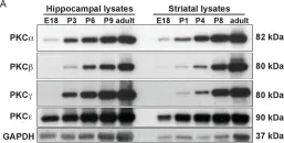
Loss of epithelial integrity is associated with colorectal cancer (CRC) aggressiveness. Protein kinase C (PKC) is frequently implicated in human cancers, but the role of PKCγ in CRC remains poorly understood. Here, we show that PKCγ, a conventional PKC, is expressed in normal colonic epithelium, but this is lower in dedifferentiated CRC. PKCγ expression was downregulated by SNAI1 overexpression, and low PKCγ expression was associated with poor prognosis in patients with CRC. Transient or stable knockdown of PKCγ reduced E-cadherin expression in CRC cells. PKCγ knockdown enhanced proliferation, anchorage-independent cell growth, resistance to anti-cancer drugs, and in vivo tumor growth of DLD-1 cells. We have also identified phosphorylation substrates for PKCγ. Among them, ARHGEF18, a RhoA activator that stabilizes cell-cell junctions, was phosphorylated and stabilized by PKCγ. Thus, these results suggest that the downregulation of PKCγ decreases the epithelial property of CRC cells and enhances its malignant phenotypes.
© 2022 The Authors.
-
Homo sapiens (Human)
-
Cancer Research
In Elife on 7 November 2017 by Carriba, P. & Davies, A. M.
Fig.6.A

-
WB
-
Collected and cropped from Elife by CiteAb, provided under a CC-BY license
Image 1 of 2
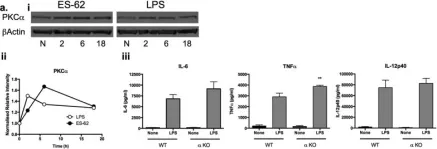
In Sci Rep on 21 November 2016 by Eason, R. J., Bell, K. S., et al.
Fig.3.A

-
WB
-
Mus musculus (House mouse)
Collected and cropped from Sci Rep by CiteAb, provided under a CC-BY license
Image 1 of 2