Central areolar choroidal dystrophy is an inherited disorder characterized by progressive choriocapillaris atrophy and retinal degeneration and is usually associated with mutations in the PRPH2 gene. We aimed to generate and characterize a mouse model with the p.Arg195Leu mutation previously described in patients. Heterozygous (Prph2WT/KI) and homozygous (Prph2KI/KI) mice were generated using the CRISPR/Cas9 system to introduce the p.Arg195Leu mutation. Retinal function was assessed by electroretinography and optomotor tests at 1, 3, 6, 9, 12, and 20 months of age. The structural integrity of the retinas was evaluated at the same ages using optical coherence tomography. Immunofluorescence and transmission electron microscopy images of the retina were also analyzed. Genetic sequencing confirmed that both Prph2WT/KI and Prph2KI/KI mice presented the p.Arg195Leu mutation. A progressive loss of retinal function was found in both mutant groups, with significantly reduced visual acuity from 3 months of age in Prph2KI/KI mice and from 6 months of age in Prph2WT/KI mice. Decreased amplitudes in the electroretinography responses were observed from 1 month of age in Prph2KI/KI mice and from 6 months of age in Prph2WT/KI mice. Morphological analysis of the retinas correlated with functional findings, showing a progressive decrease in retinal thickness of mutant mice, with earlier and more severe changes in the homozygous mutant mice. We corroborated the alteration of the outer segment structure, and we found changes in the synaptic connectivity in the outer plexiform layer as well as gliosis and signs of microglial activation. The new Prph2WT/KI and Prph2KI/KI murine models show a pattern of retinal degeneration similar to that described in human patients with central areolar choroidal dystrophy and appear to be good models to study the mechanisms involved in the onset and progression of the disease, as well as to test the efficacy of new therapeutic strategies.
© 2023. The Author(s).
Product Citations: 33
In Cell Death & Disease on 1 November 2023 by Ruiz-Pastor, M. J., Sánchez-Sáez, X., et al.
-
Mus musculus (House mouse)
-
Cell Biology
-
Neuroscience
Preprint on Research Square on 10 August 2023 by Belsamma, L. V., Muraleedharan, A., et al.
Breast cancer is the leading cause of cancer-related deaths in women, with metastasis being the primary reason for mortality. Patients with triple-negative breast cancer (TNBC) show an increased risk of metastatic dissemination. Protein kinase C eta (PKCη), an anti-apoptotic kinase of the novel PKC subfamily, is associated with poor prognosis in breast cancer patients. Here, we demonstrate that PKCη promotes metastasis in TNBC cells and show that this is mediated by the PKCƞ-YAP signaling axis. Knockout of PKCη (PKCη KO ) in the TNBC cells, 4T1 and MDA-MB-231, markedly inhibited their invasion and migration capability. Furthermore, orthotopic xenografts of the latter PKCη KO cells in NSG mice reduced tumor growth and lung metastasis compared to PKCη-intact tumors. Mechanistically, we show that PKCη regulates epithelial-to-mesenchymal transition (EMT), as knockout of PKCη in TNBC cell lines increased expression of the EMT markers E-cadherin, EpCAM, and slug, and decreased expression of vimentin, ZEB1. Further profiling of the Hippo-YAP axis showed that PKCη is a negative regulator of the Hippo pathway that leads to YAP stabilization and its phosphorylation at Ser128, which allows YAP to translocate to the nucleus and contribute to metastasis of TNBC cells. We further show that PKCη directly interacts with YAP in silico and TNBC cells. Lastly, we demonstrate that treatment of TNBC cells with uPEP2, a recently discovered PKCη kinase inhibitory peptide (encoded by a uORF upstream of PKCη coding sequence), activates the Hippo pathway and YAP degradation. In summary, our results highlight the impact of PKCη in TNBC metastasis and offer a novel avenue for therapeutic intervention in this aggressive and fatal disease.
-
Cancer Research
Immunoproteasome Inhibition Ameliorates Aged Dystrophic Mouse Muscle Environment.
In International Journal of Molecular Sciences on 24 November 2022 by Tripodi, L., Molinaro, D., et al.
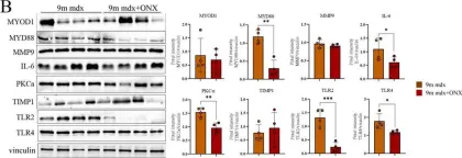
Muscle wasting is a major pathological feature observed in Duchenne muscular dystrophy (DMD) and is the result of the concerted effects of inflammation, oxidative stress and cell senescence. The inducible form of proteasome, or immunoproteasome (IP), is involved in all the above mentioned processes, regulating antigen presentation, cytokine production and immune cell response. IP inhibition has been previously shown to dampen the altered molecular, histological and functional features of 3-month-old mdx mice, the animal model for DMD. In this study, we described the role of ONX-0914, a selective inhibitor of the PSMB8 subunit of immunoproteasome, in ameliorating the pathological traits that could promote muscle wasting progression in older, 9-month-old mdx mice. ONX-0914 reduces the number of macrophages and effector memory T cells in muscle and spleen, while increasing the number of regulatory T cells. It modulates inflammatory markers both in skeletal and cardiac muscle, possibly counteracting heart remodeling and hypertrophy. Moreover, it buffers oxidative stress by improving mitochondrial efficiency. These changes ultimately lead to a marked decrease of fibrosis and, potentially, to more controlled myofiber degeneration/regeneration cycles. Therefore, ONX-0914 is a promising molecule that may slow down muscle mass loss, with relatively low side effects, in dystrophic patients with moderate to advanced disease.
-
WB
-
Mus musculus (House mouse)
In Nature Communications on 23 November 2022 by Lordén, G., Wozniak, J. M., et al.
Exquisitely tuned activity of protein kinase C (PKC) isozymes is essential to maintaining cellular homeostasis. Whereas loss-of-function mutations are generally associated with cancer, gain-of-function variants in one isozyme, PKCα, are associated with Alzheimer's disease (AD). Here we show that the enhanced activity of one variant, PKCα M489V, is sufficient to rewire the brain phosphoproteome, drive synaptic degeneration, and impair cognition in a mouse model. This variant causes a modest 30% increase in catalytic activity without altering on/off activation dynamics or stability, underscoring that enhanced catalytic activity is sufficient to drive the biochemical, cellular, and ultimately cognitive effects observed. Analysis of hippocampal neurons from PKCα M489V mice reveals enhanced amyloid-β-induced synaptic depression and reduced spine density compared to wild-type mice. Behavioral studies reveal that this mutation alone is sufficient to impair cognition, and, when coupled to a mouse model of AD, further accelerates cognitive decline. The druggability of protein kinases positions PKCα as a promising therapeutic target in AD.
© 2022. The Author(s).
-
Neuroscience
In Cell Death & Disease on 19 November 2022 by Farini, A., Tripodi, L., et al.
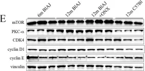
Muscle repair in dysferlinopathies is defective. Although macrophage (Mø)-rich infiltrates are prominent in damaged skeletal muscles of patients with dysferlinopathy, the contribution of the immune system to the disease pathology remains to be fully explored. Numbers of both pro-inflammatory M1 Mø and effector T cells are increased in muscle of dysferlin-deficient BlAJ mice. In addition, symptomatic BlAJ mice have increased muscle production of immunoproteasome. In vitro analyses using bone marrow-derived Mø of BlAJ mice show that immunoproteasome inhibition results in C3aR1 and C5aR1 downregulation and upregulation of M2-associated signaling. Administration of immunoproteasome inhibitor ONX-0914 to BlAJ mice rescues muscle function by reducing muscle infiltrates and fibro-adipogenesis. These findings reveal an important role of immunoproteasome in the progression of muscular dystrophy in BlAJ mouse and suggest that inhibition of immunoproteasome may produce therapeutic benefit in dysferlinopathy.
© 2022. The Author(s).
-
WB
-
Mus musculus (House mouse)
-
Cell Biology
-
Immunology and Microbiology
-
Pathology
In Int J Mol Sci on 24 November 2022 by Tripodi, L., Molinaro, D., et al.
Fig.4.B

-
WB
-
Collected and cropped from Int J Mol Sci by CiteAb, provided under a CC-BY license
Image 1 of 4
In Cell Death Dis on 19 November 2022 by Farini, A., Tripodi, L., et al.
Fig.7.E

-
WB
-
Mus musculus (House mouse)
Collected and cropped from Cell Death Dis by CiteAb, provided under a CC-BY license
Image 1 of 4
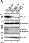
In J Biol Chem on 23 February 2021 by Van, A. N., Kunkel, M. T., et al.
Fig.5.A

-
WB
-
Collected and cropped from J Biol Chem by CiteAb, provided under a CC-BY license
Image 1 of 4
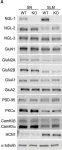
In Front Mol Neurosci on 4 June 2019 by Choi, Y., Park, H., et al.
Fig.7.A

-
WB
-
Mus musculus (House mouse)
Collected and cropped from Front Mol Neurosci by CiteAb, provided under a CC-BY license
Image 1 of 4