Lethal toxin (LT) is the critical virulence factor of Bacillus anthracis, the causative agent of anthrax. One common symptom observed in patients with anthrax is thrombocytopenia, which has also been observed in mice injected with LT. Our previous study demonstrated that LT induces thrombocytopenia by suppressing megakaryopoiesis, but the precise molecular mechanisms behind this phenomenon remain unknown. In this study, we utilized 12-O-tetradecanoylphorbol-13-acetate (TPA)-induced megakaryocytic differentiation in human erythroleukemia (HEL) cells to identify genes involved in LT-induced megakaryocytic suppression. Through cDNA microarray analysis, we identified Dachshund homolog 1 (DACH1) as a gene that was upregulated upon TPA treatment but downregulated in the presence of TPA and LT, purified from the culture supernatants of B. anthracis. To investigate the function of DACH1 in megakaryocytic differentiation, we employed short hairpin RNA technology to knock down DACH1 expression in HEL cells and assessed its effect on differentiation. Our data revealed that the knockdown of DACH1 expression suppressed megakaryocytic differentiation, particularly in polyploidization. We demonstrated that one mechanism by which B. anthracis LT induces suppression of polyploidization in HEL cells is through the cleavage of MEK1/2. This cleavage results in the downregulation of the ERK signaling pathway, thereby suppressing DACH1 gene expression and inhibiting polyploidization. Additionally, we found that known megakaryopoiesis-related genes, such as FOSB, ZFP36L1, RUNX1, FLI1, AHR, and GFI1B genes may be positively regulated by DACH1. Furthermore, we observed an upregulation of DACH1 during in vitro differentiation of CD34-megakaryocytes and downregulation of DACH1 in patients with thrombocytopenia. In summary, our findings shed light on one of the molecular mechanisms behind LT-induced thrombocytopenia and unveil a previously unknown role for DACH1 in megakaryopoiesis.
Product Citations: 58
In International Journal of Molecular Sciences on 7 March 2024 by Lin, G. L., Chang, H. H., et al.
In European Journal of Immunology on 1 March 2024 by Alsouri, S., Ambrose, A., et al.
The structure and dynamics of F-actin networks in the cortical area of B cells control the signal efficiency of B-cell antigen receptors (BCRs). Although antigen-induced signaling has been studied extensively, the role of cortical F-actin in antigen-independent tonic BCR signaling is less well understood. Because these signals are essential for the survival of B cells and are consequently exploited by several B-cell lymphomas, we assessed how the cortical F-actin structure influences tonic BCR signal transduction. We employed genetic variants of a primary cell-like B-cell line that can be rendered quiescent to show that cross-linking of actin filaments by α-actinin-4 (ACTN4), but not ACTN1, is required to preserve the dense architecture of F-actin in the cortical area of B cells. The reduced cortical F-actin density in the absence of ACTN4 resulted in increased lateral BCR diffusion. Surprisingly, this was associated with reduced tonic activation of BCR-proximal effector proteins, extracellular signal-regulated kinase, and pro-survival pathways. Accordingly, ACTN4-deficient B-cell lines and primary human B cells exhibit augmented apoptosis. Hence, our findings reveal that cortical F-actin architecture regulates antigen-independent tonic BCR survival signals in human B cells.
© 2024 Wiley-VCH GmbH.
-
Immunology and Microbiology
Overcoming MET-mediated resistance in oncogene-driven NSCLC.
In IScience on 21 July 2023 by Reischmann, N., Schmelas, C., et al.
This study evaluates the efficacy of combining targeted therapies with MET or SHP2 inhibitors to overcome MET-mediated resistance in different NSCLC subtypes. A prevalence study was conducted for MET amplification and overexpression in samples from patients with NSCLC who relapsed on ALK, ROS1, or RET tyrosine kinase inhibitors. MET-mediated resistance was detected in 37.5% of tissue biopsies, which allow the detection of MET overexpression, compared to 7.4% of liquid biopsies. The development of drug resistance by MET overexpression was confirmed in EGFRex19del-, KRASG12C-, HER2ex20ins-, and TPM3-NTRK1-mutant cell lines. The combination of targeted therapy with MET or SHP2 inhibitors was found to overcome MET-mediated resistance in both in vitro and in vivo assays. This study highlights the importance of considering MET overexpression as a resistance driver to NSCLC targeted therapies to better identify patients who could potentially benefit from combination approaches with MET or SHP2 inhibitors.
© 2023 The Authors.
In International Journal of Molecular Sciences on 30 December 2022 by Sharma, D., Rasool, F., et al.
Cancer is one of the leading cause of lethality worldwide, CRC being the third most common cancer reported worldwide, with 1.85 million cases and 850,000 deaths annually. As in all other cancers, kinases are one of the major enzymes that play an essential role in the incidence and progression of CRC. Thus, using multi-kinase inhibitors is one of the therapeutic strategies used to counter advanced-stage CRC. Regorafenib is an FDA-approved drug in the third-line therapy of refractory metastatic colorectal cancer. Acquired resistance to cancers and higher toxicity of these drugs are disadvantages to the patients. To counter this, combination therapy is used as a strategy where a minimal dose of drugs can be used to get a higher efficacy and reduce drug resistance development. Ruthenium-based compounds are observed to be a potential alternative to platinum-based drugs due to their significant safety and effectiveness. Formerly, our lab reported Ru-1, a ruthenium-based compound, for its anticancer activity against multiple cancer cells, such as HepG2, HCT116, and MCF7. This study evaluates Ru-1's activity against regorafenib-resistant HCT116 cells and as a combination therapeutic with regorafenib. Meanwhile, the mechanism of the effect of Ru-1 alone and with regorafenib as a combination is still unknown. In this study, we tested a drug combination (Ru-1 and regorafenib) against a panel of HT29, HCT116, and regorafenib-resistant HCT116 cells. The combination showed a synergistic inhibitory activity. Several mechanisms underlying these numerous synergistic activities, such as anti-proliferative efficacy, indicated that the combination exhibited potent cytotoxicity and enhanced apoptosis induction. Disruption of mitochondrial membrane potential increased intracellular ROS levels and decreased migratory cell properties were observed. The combination exhibited its activity by regulating PI3K/Akt and p38 MAP kinase signalling. This indicates that the combination of REG/Ru-1 targets cancer cells by modulating the PI3K/Akt and ERK signalling.
-
WB
-
Cancer Research
In Developmental Cell on 10 October 2022 by Hino, N., Matsuda, K., et al.
Upon the initiation of collective cell migration, the cells at the free edge are specified as leader cells; however, the mechanism underlying the leader cell specification remains elusive. Here, we show that lamellipodial extension after the release from mechanical confinement causes sustained extracellular signal-regulated kinase (ERK) activation and underlies the leader cell specification. Live-imaging of Madin-Darby canine kidney (MDCK) cells and mouse epidermis through the use of Förster resonance energy transfer (FRET)-based biosensors showed that leader cells exhibit sustained ERK activation in a hepatocyte growth factor (HGF)-dependent manner. Meanwhile, follower cells exhibit oscillatory ERK activation waves in an epidermal growth factor (EGF) signaling-dependent manner. Lamellipodial extension at the free edge increases the cellular sensitivity to HGF. The HGF-dependent ERK activation, in turn, promotes lamellipodial extension, thereby forming a positive feedback loop between cell extension and ERK activation and specifying the cells at the free edge as the leader cells. Our findings show that the integration of physical and biochemical cues underlies the leader cell specification during collective cell migration.
Copyright © 2022 Elsevier Inc. All rights reserved.
-
WB
-
Canis lupus familiaris (Domestic dog)
-
Stem Cells and Developmental Biology
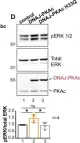
In Elife on 7 May 2019 by Turnham, R. E., Smith, F. D., et al.
Fig.6.D

-
WB
-
Homo sapiens (Human)
Collected and cropped from Elife by CiteAb, provided under a CC-BY license
Image 1 of 5
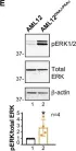
In Elife on 7 May 2019 by Turnham, R. E., Smith, F. D., et al.
Fig.5.E

-
WB
-
Homo sapiens (Human)
Collected and cropped from Elife by CiteAb, provided under a CC-BY license
Image 1 of 5
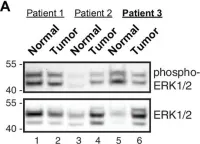
In Elife on 7 May 2019 by Turnham, R. E., Smith, F. D., et al.
Fig.4.A

-
WB
-
Homo sapiens (Human)
Collected and cropped from Elife by CiteAb, provided under a CC-BY license
Image 1 of 5
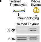
In iScience on 21 December 2018 by Konishi, Y., Terai, K., et al.
Fig.3.H

-
WB
-
Collected and cropped from iScience by CiteAb, provided under a CC-BY license
Image 1 of 5
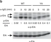
In Nat Commun on 21 November 2014 by Engels, N., König, L. M., et al.
Fig.8.B

-
WB
-
Collected and cropped from Nat Commun by CiteAb, provided under a CC-BY license
Image 1 of 5