Oxidative stress-caused damage to the retinal pigment epithelium (RPE) underlies the onset and progression of age-related macular degeneration (AMD). Impaired mitochondrial biogenesis sensitizes RPE cells to mitochondrial dysfunction, energy insufficiency and death. Src-homology 2 domain-containing phosphatase (SHP)-1 is important in regulating immune responses and cell survival. However, its roles in cell survival are not always consistent. Until now, the effects of SHP-1 on RPE dysfunction, especially mitochondrial homeostasis, remain to be elucidated. We sought to clarify the effects of SHP-1 in RPE cells in response to atRAL-induced oxidative stress and determine the regulatory mechanisms involved.
In the all trans retinal (atRAL)-induced oxidative stress model, we used the vector of lentivirus to knockdown the expression of SHP-1 in ARPE-19 cells. CCK-8 assay, Annexin V/PI staining and JC-1 staining were utilized to determine the cell viability, cell apoptosis and mitochondrial membrane potential. We also used immunoprecipitation to examine the ubiquitination modification of stimulator of interferon genes (STING) and its interaction with SHP-1. The expression levels of mitochondrial marker, proteins related to mitochondrial biogenesis, and signaling molecules involved were examined by western blotting analysis.
We found that SHP-1 knockdown predisposed RPE cells to apoptosis, aggravated mitochondrial damage, and repressed mitochondrial biogenesis after treatment with atRAL. Immunofluoresent staining and immunoprecipitation analysis confirmed that SHP-1 interacted with the endoplasmic reticulum-resident STING and suppressed K63-linked ubiquitination and activation of STING. Inhibition of STING with the specific antagonist H151 attenuated the effects of SHP-1 knockdown on mitochondrial biogenesis and oxidative damage. The adenosine monophosphate-activated protein kinase (AMPK) pathway acted as the crucial downstream target of STING and was involved in the regulatory processes.
These findings suggest that SHP-1 knockdown potentiates STING overactivation and represses mitochondrial biogenesis and cell survival, at least in part by blocking the AMPK pathway in RPE cells. Therefore, restoring mitochondrial health by regulating SHP-1 in RPE cells may be a potential therapeutic strategy for degenerative retinal diseases including AMD.
© 2022. The Author(s).
Product Citations: 5
In Molecular Medicine on 22 October 2022 by Zhuang, X., Ma, J., et al.
-
IHC
-
Mus musculus (House mouse)
-
Biochemistry and Molecular biology
-
Cell Biology
In Cell Death Discovery on 7 March 2020 by Wei, H., Davies, J. E., et al.
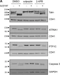
Activated, procoagulant platelets shed phosphatidylserine (PS)-exposing extracellular vesicles (EVs) from their surface in a Ca2+- and calpain-dependent manner. These PS-exposing EVs are prothrombotic and proinflammatory and are found at elevated levels in many cardiovascular and metabolic diseases. How PS-exposing EVs are shed is not fully understood. A clearer understanding of this process may aid the development of drugs to selectively block their release. In this study we report that 2-aminoethoxydiphenylborate (2-APB) significantly inhibits the release of PS-exposing EVs from platelets stimulated with the Ca2+ ionophore, A23187, or the pore-forming toxin, streptolysin-O. Two analogues of 2-APB, diphenylboronic anhydride (DPBA) and 3-(diphenylphosphino)-1-propylamine (DP3A), inhibited PS-exposing EV release with similar potency. Although 2-APB and DPBA weakly inhibited platelet PS exposure and calpain activity, this was not seen with DP3A despite inhibiting PS-exposing EV release. These data suggest that there is a further target of 2-APB, independent of cytosolic Ca2+ signalling, PS exposure and calpain activity, that is required for PS-exposing EV release. DP3A is likely to inhibit the same target, without these other effects. Identifying the target of 2-APB, DPBA and DP3A may provide a new way to inhibit PS-exposing EV release from activated platelets and inhibit their contribution to thrombosis and inflammation.
© The Author(s) 2020.
-
WB
-
Homo sapiens (Human)
In Physiological Reports on 1 April 2019 by Wang, H., Morris, R. G., et al.
Sickle cell disease (SCD)-induced urinary concentration defect has been proposed as caused by impaired ability of the occluded vasa recta due to red blood cell sickling to serve as countercurrent exchangers and renal tubules to absorb water and solutes. However, the exact molecular mechanisms remain largely unknown. The present studies were undertaken to determine the effects of SCD on vasopressin, aquaporin2 (AQP2), urea transporter A1 (UTA1), Na-K-Cl co-transporter 2 (NKCC2), epithelial Na channels (ENaC), aquaporin1 (AQP1), nuclear factor of activated T cells 5 (NFAT5) and Src homology region-2 domain-containing phosphatase-1 (SHP-1), an important regulator of NFAT5, in the Berkeley SCD mouse kidney medulla. Under water repletion, SCD only induced a minor urinary concentration defect associated with increased urinary vasopressin level alone with the well-known effects of vasopressin: protein abundance of AQP2, UTA1 and ENaC-β and apical targeting of AQP2 as compared with non-SCD. SCD did not significantly affect AQP1 protein level. Water restriction had no further significant effect on SCD urinary vasopressin. NFAT5 is also critical to urinary concentration. Instead, water restriction-activated NFAT5 associated with inhibition of SHP-1 in the SCD mice. Yet, water restriction only elevated urinary osmolality by 28% in these mice as opposed to 104% in non-SCD mice despite similar degree increases of protein abundance of AQP2, NKCC2 and AQP2-S256-P. Water-restriction had no significant effect on protein abundance of ENaC or AQP1 in either strain. In conclusion, under water repletion SCD, only induces a minor defect in urinary concentration because of compensation from the up-regulated vasopressin system. However, under water restriction, SCD mice struggle to concentrate urine despite activating NFAT5. SCD-induced urinary concentration defect appears to be resulted from the poor blood flow in vasa recta rather than the renal tubules' ability to absorb water and solutes.
© Published 2019. This article is a U.S. Government work and is in the public domain in the USA. Physiological Reports published by Wiley Periodicals, Inc. on behalf of The Physiological Society and the American Physiological Society.
-
WB
-
Endocrinology and Physiology
-
Neuroscience
In Cell Research on 1 August 2015 by Qian, H., Deng, X., et al.
Hepatocytes are critical for the maintenance of liver homeostasis, but its involvement in hepatic fibrogenesis remains elusive. Hepatocyte nuclear factor 1α (HNF1α) is a liver-enriched transcription factor that plays a key role in hepatocyte function. Our previous study revealed a significant inhibitory effect of HNF1α on hepatocellular carcinoma. In this study, we report that the expression of HNF1α is significantly repressed in both human and rat fibrotic liver. Knockdown of HNF1α in the liver significantly aggravates hepatic fibrogenesis in either dimethylnitrosamine (DMN) or bile duct ligation (BDL) model in rats. In contrast, forced expression of HNF1α markedly alleviates hepatic fibrosis. HNF1α regulates the transcriptional expression of SH2 domain-containing phosphatase-1 (SHP-1) via directly binding to SHP-1 promoter in hepatocytes. Inhibition of SHP-1 expression abrogates the anti-fibrotic effect of HNF1α in DMN-treated rats. Moreover, HNF1α repression in primary hepatocytes leads to the activation of NF-κB and JAK/STAT pathways and initiates an inflammatory feedback circuit consisting of HNF1α, SHP-1, STAT3, p65, miR-21 and miR-146a, which sustains the deregulation of HNF1α in hepatocytes. More interestingly, a coordinated crosstalk between hepatocytes and hepatic stellate cells (HSCs) participates in this positive feedback circuit and facilitates the progression of hepatocellular damage. Our findings demonstrate that impaired hepatocytes play an active role in hepatic fibrogenesis. Early intervention of HNF1α-regulated inflammatory feedback loop in hepatocytes may have beneficial effects in the treatment of chronic liver diseases.
-
WB
-
Cell Biology
Endotoxin-induced growth hormone resistance in skeletal muscle.
In Endocrinology on 1 August 2009 by Chen, Y., Sood, S., et al.
Inflammation-induced skeletal muscle wasting is a serious clinical problem and arises in part because of resistance to GH-stimulated IGF-I expression. Although it is established that in the liver, resistance develops because of impaired signaling through the Janus kinase 2 (JAK2)/signal transducer and activator of transcription 5 (STAT5) transduction pathway, together with a more distal defect in STAT5 DNA-binding activity, the situation in skeletal muscle is unclear. Accordingly, we set out to characterize the mechanisms behind the skeletal muscle resistance to GH in rats with acute inflammation induced by endotoxin. Endotoxin caused significant declines in GH-stimulated STAT5a/b phosphorylation and IGF-I gene expression, and this occurred despite a lack of change in signaling protein levels or phosphorylation of JAK2. In whole muscle, GH-stimulated phospho-STAT5a/b levels were reduced by half, and in the nucleus, phospho-STAT5b levels were similarly reduced. Furthermore, the binding of phosphorylated STAT5b to DNA was reduced and to a similar extent to the reduction in nuclear phosphorylated STAT5b. Interestingly, GH-induced androgen receptor gene expression was also suppressed. Thus, it appears that skeletal muscle resistance to GH-stimulated IGF-I expression in acute endotoxemia arises from a defect in STAT5b signaling, with a proportionate reduction in STAT5b DNA binding. Finally, it appears that resistance to GH-induced androgen receptor expression also develops and, together with the attenuated GH-induced IGF-I expression, likely plays an important role in the muscle wasting that arises in endotoxin-induced inflammation.
-
Endocrinology and Physiology
In Cell Death Discov on 7 March 2020 by Wei, H., Davies, J. E., et al.
Fig.6.A

-
WB
-
Homo sapiens (Human)
Collected and cropped from Cell Death Discov by CiteAb, provided under a CC-BY license
Image 1 of 1