Pneumolysin (PLY) is a bacterial pore forming toxin and primary virulence factor of Streptococcus pneumonia, a major cause of pneumonia. PLY binds cholesterol-rich domains of the endothelial cell (EC) plasma membrane resulting in pore assembly and increased intracellular (IC) Ca2+ levels that compromise endothelial barrier integrity. Caveolae are specialized plasmalemma microdomains of ECs enriched in cholesterol. We hypothesized that the abundance of cholesterol-rich domains in EC plasma membranes confers cellular susceptibility to PLY. Contrary to this hypothesis, we found increased PLY-induced IC Ca2+ following membrane cholesterol depletion. Caveolin-1 (Cav-1) is an essential structural protein of caveolae and its regulation by cholesterol levels suggested a possible role in EC barrier function. Indeed, Cav-1 and its scaffolding domain peptide protected the endothelial barrier from PLY-induced disruption. In loss of function experiments, Cav-1 was knocked-out using CRISPR-Cas9 or silenced in human lung microvascular ECs. Loss of Cav-1 significantly enhanced the ability of PLY to disrupt endothelial barrier integrity. Rescue experiments with re-expression of Cav-1 or its scaffolding domain peptide protected the EC barrier against PLY-induced barrier disruption. Dynamin-2 (DNM2) is known to regulate caveolar membrane endocytosis. Inhibition of endocytosis, with dynamin inhibitors or siDNM2 amplified PLY induced EC barrier dysfunction. These results suggest that Cav-1 protects the endothelial barrier against PLY by promoting endocytosis of damaged membrane, thus reducing calcium entry and PLY-dependent signaling.
Copyright © 2022 Batori, Chen, Bordan, Haigh, Su, Verin, Barman, Stepp, Chakraborty, Lucas and Fulton.
Product Citations: 11
Protective role of Cav-1 in pneumolysin-induced endothelial barrier dysfunction.
In Frontiers in Immunology on 16 August 2022 by Batori, R. K., Chen, F., et al.
-
WB
-
Immunology and Microbiology
Dynamics and nanoscale organization of the postsynaptic endocytic zone at excitatory synapses.
In eLife on 24 January 2022 by Catsburg, L. A. E., Westra, M., et al.
At postsynaptic sites of neurons, a prominent clathrin-coated structure, the endocytic zone (EZ), controls the trafficking of glutamate receptors and is essential for synaptic plasticity. Despite its importance, little is known about how this clathrin structure is organized to mediate endocytosis. We used live-cell and super-resolution microscopy to reveal the dynamic organization of this poorly understood clathrin structure in rat hippocampal neurons. We found that a subset of endocytic proteins only transiently appeared at postsynaptic sites. In contrast, other proteins were persistently enriched and partitioned at the edge of the EZ. We found that uncoupling the EZ from the synapse led to the loss of most of these components, while disrupting interactions with the actin cytoskeleton or membrane did not alter EZ positioning. Finally, we found that plasticity-inducing stimuli promoted the reorganization of the EZ. We conclude that the EZ is a stable, highly organized molecular platform where components are differentially recruited and positioned to orchestrate the endocytosis of synaptic receptors.
© 2022, Catsburg et al.
-
ICC
-
Rattus norvegicus (Rat)
-
Neuroscience
In Communications Biology on 13 March 2020 by Cao, F., Zhou, Y., et al.
Integrin receptors orchestrate cell adhesion and cytoskeletal reorganization. The endocytic mechanism of integrin-β3 receptor at the podosome remains unclear. Using viscous RGD-membrane as the model system, here we show that the formation of podosome-like adhesion promotes Dab2/clathrin-mediated endocytosis of integrin-β3. Integrin-β3 and RGD ligand are endocytosed from the podosome and sorted into the endosomal compartment. Inhibitions of podosome formation and knockdowns of Dab2 and clathrin reduce RGD endocytosis. F-actin assembly at the podosome core exhibits protrusive contact towards the substrate and results in plasma membrane invaginations at the podosome ring. BIN1 specifically associates with the region of invaginated membrane and recruits DNM2. During the podosome formation, BIN1 and DNM2 synchronously enrich at the podosome ring and trigger clathrin dissociation and RGD endocytosis. Knockdowns of BIN1 and DNM2 suppress RGD endocytosis. Thus, plasma membrane invagination caused by F-actin polymerization promotes BIN1-dependent DNM2 recruitment and facilitate integrin-β3 endocytosis at the podosome.
-
WB
In Scientific Reports on 11 January 2019 by Yotsuya, M., Bertagna, A. E., et al.
The degeneration of articular cartilage underscores the clinical pathology of temporomandibular joint osteoarthritis (TMJ-OA) and is promoted through dysfunctional biochemical or biophysical signaling. Transduction of these signals has a multifaceted regulation that includes important cell-matrix derived interactions. The matrix encapsulating the cells of the mandibular condylar cartilage (MCC) is rich in type VI collagen. Neuron/glia antigen 2 (NG2) is a type I transmembrane proteoglycan that binds with type VI collagen. This study defines the temporospatial dynamics of NG2-type VI collagen interactions during the progression of TMJ-OA. Membrane-bound NG2 is found to colocalize with pericellular type VI collagen in superficial layer cells in the MCC perichondrium but is present at high levels in the cytosol of chondroblastic and hypertrophic cells. When TMJ -OA is induced using a surgical instability model, localized disruptions of pericellular type VI collagen are observed on the central and medial MCC and are associated with significantly higher levels of cytosolic NG2. NG2 localized within the cytosol is found to be transported through clathrin and dynamin mediated endocytic pathways. These findings are consistent with NG2 behavior in other injury models and underscore the potential of NG2 as an entirely novel molecular mechanism of chondrocyte function contextually linked with TMJ-OA.
-
IHC
-
Mus musculus (House mouse)
-
Immunology and Microbiology
-
Neuroscience
In BMC Neuroscience on 18 July 2016 by Thomas, R. S., Henson, A., et al.
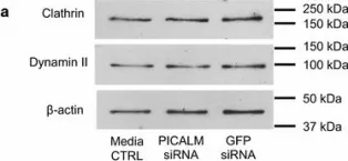
Polymorphisms in the gene for phosphatidylinositol binding clathrin assembly protein (PICALM), an endocytic-related protein, are associated with a small, increased risk of developing Alzheimer's disease (AD), strongly suggesting that changes in endocytosis are involved in the aetiology of the disease. We have investigated the involvement of PICALM in the processing of amyloid precursor protein (APP) to understand how PICALM could be linked to the development of AD. We used siRNA to deplete levels of PICALM, its isoforms and clathrin heavy chain in the human brain-derived H4 neuroglioma cell line that expresses endogenous levels of APP. We then used Western blotting, ELISA and immunohistochemistry to detect intra- and extracellular protein levels of endocytic-related proteins, APP and APP metabolites including β-amyloid (Aβ). Levels of functional endocytosis were quantified using ALEXA 488-conjugated transferrin and flow cytometry as a marker of clathrin-mediated endocytosis (CME).
Following depletion of all the isoforms of PICALM by siRNA in H4 cells, levels of intracellular APP, intracellular β-C-terminal fragment (β-CTF) and secreted sAPPβ (APP fragments produced by β-secretase cleavage) were significantly reduced but Aβ40 was not affected. Functional endocytosis was significantly reduced after both PICALM and clathrin depletion, highlighting the importance of PICALM in this process. However, depletion of clathrin did not affect APP but did reduce β-CTF levels. PICALM depletion altered the intracellular distribution of clathrin while clathrin reduction affected the subcellular pattern of PICALM labelling. Both PICALM and clathrin depletion reduced the expression of BACE1 mRNA and PICALM siRNA reduced protein levels. Individual depletion of PICALM isoforms 1 and 2 did not affect APP levels while clathrin depletion had a differential effect on the isoforms, increasing isoform 1 while decreasing isoform 2 expression.
The depletion of PICALM in brain-derived cells has significant effects on the processing of APP, probably by reducing CME. In particular, it affects the production of β-CTF which is increasingly considered to be an important mediator in AD independent of Aβ. Thus a decrease in PICALM expression in the brain could be beneficial to slow or prevent the development of AD.
-
WB
-
Neuroscience
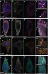
In Sci Rep on 11 January 2019 by Yotsuya, M., Bertagna, A. E., et al.
Fig.5.A

-
IHC
-
Mus musculus (House mouse)
Collected and cropped from Sci Rep by CiteAb, provided under a CC-BY license
Image 1 of 3
In BMC Neurosci on 18 July 2016 by Thomas, R. S., Henson, A., et al.
Fig.3.A

-
WB
-
Collected and cropped from BMC Neurosci by CiteAb, provided under a CC-BY license
Image 1 of 3
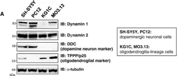
In Mol Neurodegener on 14 August 2012 by Konno, M., Hasegawa, T., et al.
Fig.4.A

-
WB
-
Collected and cropped from Mol Neurodegener by CiteAb, provided under a CC-BY license
Image 1 of 3