Although Schwann cells have been found to play a key role in inflammation and repair following nerve injury, the exact pathway is still unknown. To explore the mechanism by which Schwann cells exert their effects in the neuron microenvironment, we investigated two main inflammatory pathways: the NF-κB and cAMP pathways, and their downstream signaling molecules. In this study, lipopolysaccharide (LPS), a bacterial endotoxin, was used to activate the NF-κB pathway, and forskolin, a plant extract, was used to activate the cAMP pathway. The rat RT4-D6P2T Schwann cell line was treated with 0.1, 1, or 10 μg/mL of LPS, with or without 2 μM of forskolin, for 1, 3, 12, and 24 hours to determine the effects of elevated cAMP levels on LPS-treated cell viability. To investigate the effects of elevated cAMP levels on the expression of downstream signaling effector proteins, specifically NF-κB, TNF-α, AKAP95, and cyclin D3, as well as TNF-α secretion, RT4-D6P2T cells were incubated in the various treatment combinations for a 3-hour time period. Overall, results from the CellTiter-Glo viability assay revealed that forskolin increased viability in cells treated with smaller doses of LPS for 1 and 24 hours. For all time points, 10 μg/mL of LPS noticeably reduced viability regardless of forskolin treatment. Results from the Western blot analysis revealed that, at 10 μg/mL of LPS, forskolin upregulated the expression of TNF-α despite a downregulation of NF-κB, which was also accompanied by a decrease in TNF-α secretion. These results provide evidence that cAMP might regulate TNF-α expression through alternate pathways. Furthermore, although cAMP activation altered AKAP95 and cyclin D3 expression at different doses of LPS, there does not appear to be an association between the expression of AKAP95 or cyclin D3 and the expression of TNF-α. Exploring the possible interactions between cAMP, NF-κB, and other key inflammatory signaling pathways might reveal a potential therapeutic target for the treatment of nerve injury and inflammation.
Copyright: © 2024 Henry et al. This is an open access article distributed under the terms of the Creative Commons Attribution License, which permits unrestricted use, distribution, and reproduction in any medium, provided the original author and source are credited.
Product Citations: 16
In PLoS ONE on 16 April 2024 by Henry, C., Wilcox, M., et al.
-
Neuroscience
Combined inhibition of ACLY and CDK4/6 reduces cancer cell growth and invasion.
In Oncology Reports on 1 February 2023 by Velez, B. C., Petrella, C. P., et al.
The use of small molecule kinase inhibitors, which target specific enzymes that are overactive in cancer cells, has revolutionized cancer patient treatment. To treat some types of breast cancer, CDK4/6 inhibitors, such as palbociclib, have been developed that target the phosphorylation of the retinoblastoma tumor suppressor gene. Acquired resistance to CDK4/6 inhibitors may be due to activation of the AKT pro‑survival signaling pathway that stimulates several processes, such as growth, metastasis and changes in metabolism that support rapid cell proliferation. The aim of the present study was to investigate whether targeting ATP citrate lyase (ACLY), a downstream target of AKT, may combine with CDK4/6 inhibition to inhibit tumorigenesis. The present study determined that ACLY is activated in breast and pancreatic cancer cells in response to palbociclib treatment and AKT mediates this effect. Inhibition of ACLY using bempedoic acid used in combination with palbociclib reduced cell viability in a panel of breast and pancreatic cancer cell lines. This effect was also observed using breast cancer cells grown in 3D cell culture. Mechanistically, palbociclib inhibited cell proliferation, whereas bempedoic acid stimulated apoptosis. Finally, using Transwell invasion assays and immunoblotting, the present study demonstrated that ACLY inhibition blocked cell invasion, when used alone or in combination with palbociclib. These data may yield useful information that could guide the development of future therapies aimed at the reduction of acquired resistance observed clinically.
-
Homo sapiens (Human)
-
Cancer Research
Integrin-mediated adhesions in regulation of cellular senescence.
In Science Advances on 1 May 2020 by Shin, E. Y., Park, J. H., et al.
Bioinformatic and functional data link integrin-mediated cell adhesion to cellular senescence; however, the significance of and molecular mechanisms behind these connections are unknown. We now report that the focal adhesion-localized βPAK-interacting exchange factor (βPIX)-G protein-coupled receptor kinase interacting protein (GIT) complex controls cellular senescence in vitro and in vivo. βPIX and GIT levels decline with age. βPIX knockdown induces cellular senescence, which was prevented by reexpression. Loss of βPIX induced calpain cleavage of the endocytic adapter amphiphysin 1 to suppress clathrin-mediated endocytosis (CME); direct competition of GIT1/2 for the calpain-binding site on paxillin mediates this effect. Decreased CME and thus integrin endocytosis induced abnormal integrin signaling, with elevated reactive oxygen species production. Blocking integrin signaling inhibited senescence in human fibroblasts and mouse lungs in vivo. These results reveal a central role for integrin signaling in cellular senescence, potentially identifying a new therapeutic direction.
Copyright © 2020 The Authors, some rights reserved; exclusive licensee American Association for the Advancement of Science. No claim to original U.S. Government Works. Distributed under a Creative Commons Attribution NonCommercial License 4.0 (CC BY-NC).
In International Journal of Oncology on 1 February 2019 by Thomas, N. A., Abraham, R. G., et al.
Pancreatic ductal adenocarcinoma (PDAC) remains a particularly lethal disease that is resistant to targeted therapies. Tyrosine kinase inhibitors (TKIs), including erlotinib and gefitinib, which block the action of the human epidermal growth factor receptor type 1 receptor, provide small increases in patient survival when administered with gemcitabine. The retinoblastoma (Rb) tumor suppressor protein is an additional target in pancreatic cancer, due to its documented inactivation in PDAC. The present study, using cell number, apoptosis and immunoblotting assays, aimed to evaluate the effects of activation of the Rb tumor suppressor via dephosphorylation by small interfering RNA‑mediated phosphatase activation. In the Panc1, MIAPaCa‑2 and Capan‑2 pancreatic cancer cell lines, and in normal H6c7 cells, the effects of phosphatase activation on Rb were revealed to be dependent on expression of the p16 tumor suppressor, which regulates Rb phosphorylation. Phosphatase activation had no effect on non‑transformed pancreatic epithelial cells. When comparing kinase inhibition with phosphatase activation, it was demonstrated that kinase inhibition reduced proliferation, whereas phosphatase activation induced apoptosis. Both treatments together resulted in a greater reduction of pancreatic cancer cells than either treatment alone. In addition, the effects of combination treatment of phosphatase activation with TKIs on cell number and activation of the signal transducer and activator of transcription 3 (STAT3) resistance pathway were determined. The combination of Rb phosphatase activation with TKIs resulted in a greater reduction in cell number compared with either treatment alone, without STAT3 pathway activation. These data suggested that targeting Rb phosphorylation by activating phosphatase may be a rational strategy to inhibit pancreatic tumor cell growth, without activation of acquired resistance.
-
WB
-
Homo sapiens (Human)
-
Cancer Research
The miR-15 family reinforces the transition from proliferation to differentiation in pre-B cells.
In EMBO Reports on 1 September 2017 by Lindner, S. E., Lohmüller, M., et al.
Precursor B lymphocytes expand upon expression of a pre-B cell receptor (pre-BCR), but then transit into a resting state in which immunoglobulin light chain gene recombination is initiated. This bi-phasic sequence is orchestrated by the IL-7 receptor (IL-7R) and pre-BCR signaling, respectively, but little is known about microRNAs fine-tuning these events. Here, we show that pre-B cells lacking miR-15 family functions exhibit prolonged proliferation due to aberrant expression of the target genes cyclin E1 and D3. As a consequence, they fail to trigger the transcriptional reprogramming normally accompanying their differentiation, resulting in a developmental block at the pre-B cell stage. Intriguingly, our data indicate that the miR-15 family is suppressed by both IL-7R and pre-BCR signaling, suggesting it is actively integrated into the regulatory circuits of developing B cells. These findings identify the miR-15 family as a novel element required to promote the switch from pre-B cell proliferation to differentiation.
© 2017 The Authors.
-
WB
-
Mus musculus (House mouse)
-
Immunology and Microbiology
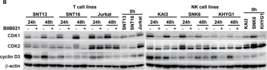
In Front Microbiol on 29 April 2015 by Suzuki, M., Takeda, T., et al.
Fig.5.B

-
WB
-
Collected and cropped from Front Microbiol by CiteAb, provided under a CC-BY license
Image 1 of 1