Secreted protein acidic and rich in cysteine (SPARC), a conserved secreted glycoprotein, plays crucial roles in regulating various biological processes. SPARC is highly expressed and has profound implications in several cancer types, including melanoma. Understanding the mechanisms that govern SPARC expression in cancers has the potential to lead to improved cancer diagnosis, prognosis, treatment strategies, and patient outcomes. Here, we demonstrate that histone deacetylase 10 (HDAC10) is a key regulator of SPARC expression in melanoma cells. Depletion or inhibition of HDAC10 upregulates SPARC expression, whereas overexpression of HDAC10 downregulates it. Mechanistically, HDAC10 coordinates with histone acetyltransferase p300 to modulate the state of acetylation of histone H3 at lysine 27 (H3K27ac) at SPARC regulatory elements and the recruitment of bromodomain-containing protein 4 (BRD4) to these regions, thereby fine-tuning SPARC transcription. HDAC10 depletion and resultant SPARC upregulation repress melanoma cell growth primarily by activating AMPK signaling and inducing autophagy. Moreover, SPARC upregulation due to HDAC10 depletion partly accounts for the resensitization of resistant cells to a BRAF inhibitor. Our work reveals the role of HDAC10 in gene regulation through indirect histone modification and suggests a potential therapeutic strategy for melanoma or other cancers by targeting HDAC10 and SPARC.
© The Author(s) 2024. Published by Oxford University Press on behalf of NAR Cancer.
Product Citations: 51
In NAR Cancer on 1 June 2024 by Ling, H., Li, Y., et al.
-
Cancer Research
Preprint on BioRxiv : the Preprint Server for Biology on 7 December 2023 by Ling, H., Li, Y., et al.
SUMMARY Secreted Protein Acidic and Rich in Cysteine (SPARC), a highly conserved secreted glycoprotein, is crucial for various bioprocesses. Here we demonstrate that histone deacetylase 10 (HDAC10) is a key regulator of SPARC expression. HDAC10 depletion or inhibition upregulates, while overexpression of HDAC10 downregulates, SPARC expression. Mechanistically, HDAC10 coordinates with histone acetyltransferase p300 to modulate the acetylation state of histone H3 lysine 27 (H3K27ac) at SPARC regulatory elements and the recruitment of bromodomain-containing protein 4 (BRD4) to these regions, thereby tuning SPARC transcription. HDAC10 depletion and resultant SPARC upregulation repress melanoma cell growth, primarily by induction of autophagy via activation of AMPK signaling. Moreover, SPARC upregulation due to HDAC10 depletion partly accounts for the resensitivity of resistant cells to a BRAF inhibitor. Our work reveals the role of HDAC10 in gene regulation through epigenetic modification and suggests a potential therapeutic strategy for melanoma or other cancers by targeting HDAC10 and SPARC. Highlights HDAC10 is the primary HDAC member that tightly controls SPARC expression. HDAC10 coordinates with p300 in modulating the H3K27ac state at SPARC regulatory elements and the recruitment of BRD4 to these regions. HDAC10 depletion and resultant SPARC upregulation inhibit melanoma cell growth by inducing autophagy via activation of AMPK signaling. SPARC upregulation as a result of HDAC10 depletion resensitizes resistant cells to BRAF inhibitors.
-
Cancer Research
In Journal of Cancer on 14 August 2023 by Kim, S., Kim, D. E., et al.
The anti-proliferative effects of a newly developed N3-acyl-N5-aryl-3,5-diaminoindazole analog, KMU-191, have been previously evaluated in various cancer cells. However, the detailed anti-cancer molecular mechanisms of KMU-191 remain unknown. In this study, we investigated anti-cancer mechanisms by which KMU-191 regulates apoptosis-related genes in human clear cell renal cell carcinoma Caki cells. KMU-191 induced poly ADP-ribose polymerase cleavage and caspase-dependent apoptosis. In addition, KMU-191 induced down-regulation of the long form of cellular FADD-like IL-1β-converting enzyme inhibitory protein (c-FLIP (L)) at the transcriptional level as well as that of long form of myeloid cell leukemia (Mcl-1 (L)) and B-cell lymphoma-extra large at the post-transcriptional level. Furthermore, KMU-191-induced apoptosis was closely associated with the Mcl-1 (L) down-regulation, but also partially associated with c-FLIP (L) down-regulation. In contrast, KMU-191 up-regulated p53, which is closely related to KMU-191-induced apoptosis. Although KMU-191 showed cytotoxicity of normal cells, it unusually did not induce cardiotoxicity. Taken together, these results suggest that a multi-target small molecule, N3-acyl-N5-aryl-3,5-diaminoindazole analog, KMU-191 is a potential anti-cancer agent that does not induce cardiotoxicity.
© The author(s).
-
Cancer Research
In Nutrients on 5 August 2022 by Park, D., Lim, J. W., et al.
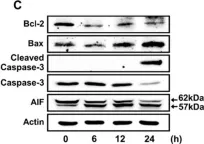
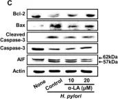
Helicobacter pylori (H. pylori) is a Gram-negative bacterium that colonizes the gastric mucosa and triggers various stomach diseases. H. pylori induces reactive oxygen species (ROS) production and DNA damage. The heterodimeric Ku70/Ku80 protein plays an essential role in the repair of DNA double-strand breaks (DSB). Oxidative stress stimulate apoptosis and DNA damage that can be repaired by Ku70/80. However, excessive reactive oxygen species (ROS) can cause Ku protein degradation, resulting in DNA fragmentation and apoptosis. α-lipoic acid (α-LA), which is found in organ meats such as liver and heart, spinach, broccoli, and potatoes, quenches free radicals, chelates metal ions, and reduces intracellular DNA damage induced by oxidative stress. Here, we investigated whether H. pylori decreases Ku70/80 and induces apoptosis, and whether α-LA inhibits changes induced by H. pylori. We analyzed ROS, DNA damage markers (γ-H2AX, DNA fragmentation), levels of Ku70/80, Ku-DNA binding activity, Ku80 ubiquitination, apoptosis indices (Bcl-2, Bax, apoptosis-inducing factor (AIF), and caspase-3), and viability in a human gastric epithelial adenocarcinoma cell line (AGS). H. pylori increased ROS, DNA damage markers, Ku80 ubiquitination, and consequently induced apoptosis. It also decreased nuclear Ku70/80 levels and Ku-DNA-binding activity; increased Bax expression, caspase-3 cleavage, and truncated AIF; but decreased Bcl-2 expression. These H. pylori-induced alterations were inhibited by α-LA. The antioxidant N-acetylcysteine and proteasome inhibitor MG-132 suppressed H. pylori-induced cell death and decreased nuclear Ku70/80 levels. The results show that oxidative stress induced Ku70/80 degradation via the ubiquitin-proteasome system, leading to its nuclear loss and apoptosis in H. pylori-infected cells. In conclusion, α-LA inhibited apoptosis induced by H. pylori by reducing ROS levels and suppressing the loss of Ku70/80 proteins in AGS cells.
-
WB
-
Immunology and Microbiology
In Biomedicine Pharmacotherapy = Biomédecine Pharmacothérapie on 1 June 2022 by Falgàs, A., García-León, A., et al.
High rates of relapsed and refractory diffuse large B-cell lymphoma (DLBCL) patients and life-threatening side effects associated with immunochemotherapy make an urgent need to develop new therapies for DLBCL patients. Immunotoxins seem very potent anticancer therapies but their use is limited because of their high toxicity. Accordingly, the self-assembling polypeptidic nanoparticle, T22-DITOX-H6, incorporating the diphtheria toxin and targeted to CXCR4 receptor, which is overexpressed in DLBCL cells, could offer a new strategy to selectively eliminate CXCR4+ DLBCL cells without adverse effects. In these terms, our work demonstrated that T22-DITOX-H6 showed high specific cytotoxicity towards CXCR4+ DLBCL cells at the low nanomolar range, which was dependent on caspase-3 cleavage, PARP activation and an increase of cells in early/late apoptosis. Repeated nanoparticle administration induced antineoplastic effect, in vivo and ex vivo, in a disseminated immunocompromised mouse model generated by intravenous injection of human luminescent CXCR4+ DLBCL cells. Moreover, T22-DITOX-H6 inhibited tumor growth in a subcutaneous immunocompetent mouse model bearing mouse CXCR4+ lymphoma cells in the absence of alterations in the hemogram, liver or kidney injury markers or on-target or off-target organ histology. Thus, T22-DITOX-H6 demonstrates a selective cytotoxicity towards CXCR4+ DLBCL cells without the induction of toxicity in non-lymphoma infiltrated organs nor hematologic toxicity.
Copyright © 2022 The Authors. Published by Elsevier Masson SAS.. All rights reserved.
-
WB
-
Homo sapiens (Human)
-
Cancer Research
In Nutrients on 5 August 2022 by Park, D., Lim, J. W., et al.
Fig.1.C

-
WB
-
Collected and cropped from Nutrients by CiteAb, provided under a CC-BY license
Image 1 of 7
In Nutrients on 5 August 2022 by Park, D., Lim, J. W., et al.
Fig.2.C

-
WB
-
Collected and cropped from Nutrients by CiteAb, provided under a CC-BY license
Image 1 of 7
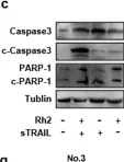
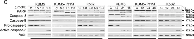
In Signal Transduct Target Ther on 24 April 2020 by Wang, Z., Liu, W., et al.
Fig.7.C

-
WB
-
Collected and cropped from Signal Transduct Target Ther by CiteAb, provided under a CC-BY license
Image 1 of 7
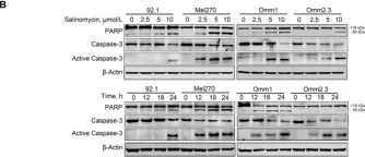
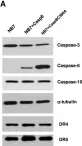
In Mol Cancer on 13 November 2019 by Zhou, J., Liu, S., et al.
Fig.2.B

-
WB
-
Collected and cropped from Mol Cancer by CiteAb, provided under a CC-BY license
Image 1 of 7
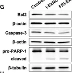
In Front Oncol on 25 July 2019 by Yu, Y., Wang, J., et al.
Fig.4.G

-
WB
-
Collected and cropped from Front Oncol by CiteAb, provided under a CC-BY license
Image 1 of 7
In Cell Death Dis on 22 January 2018 by Jin, B., Wang, C., et al.
Fig.4.C

-
WB
-
Collected and cropped from Cell Death Dis by CiteAb, provided under a CC-BY license
Image 1 of 7
In PLoS One on 23 October 2010 by Finlay, D., Richardson, R. D., et al.
Fig.1.A

-
WB
-
Homo sapiens (Human)
Collected and cropped from PLoS One by CiteAb, provided under a CC-BY license
Image 1 of 7