Extracellular vesicles (EV) have emerged as promising cell-free therapeutics in regenerative medicine. However, translating primary cell line-derived EV to clinical applications requires large-scale manufacturing and several challenges, such as replicative senescence, donor heterogeneity, and genetic instability. To address these limitations, we used a reprogramming approach to generate human induced pluripotent stem cells (hiPSC) from the young source of cord blood mesenchymal stem/stromal cells (CBMSC). Capitalizing on their inexhaustible supply potential, hiPSC offer an attractive EV reservoir. Our approach encompassed an exhaustive characterization of hiPSC-EV, aligning with the rigorous MISEV2023 guidelines. Analyses demonstrated physical features compatible with small EV (sEV) and established their identity and purity. Moreover, the sEV-shuttled non-coding (nc) RNA landscape, focusing on the microRNA and circular RNA cargo, completed the molecular signature. The kinetics of the hiPSC-sEV release and cell internalization assays unveiled robust EV production and consistent uptake by human neurons. Furthermore, hiPSC-sEV demonstrated ex vivo cell tissue-protective properties. Finally, via bioinformatics, the potential involvement of the ncRNA cargo in the hiPSC-sEV biological effects was explored. This study significantly advances the understanding of pluripotent stem cell-derived EV. We propose cord blood MSC-derived hiPSC as a promising source for potentially therapeutic sEV.
© The author(s).
Product Citations: 84
In International Journal of Biological Sciences on 12 December 2024 by Barilani, M., Peli, V., et al.
-
Stem Cells and Developmental Biology
In STAR Protocols on 21 June 2024 by Townsend, C. A., Petropavlovskiy, A. A., et al.
S-acylation, commonly palmitoylation, is the addition of fatty acids to cysteines to regulate protein localization and function. S-acylation detection has been hampered by limited sensitivity and selectivity in low-protein, costly samples like cultured neurons. Here, we present a protocol for sensitive and selective bioorthogonal labeling and click-chemistry-based detection of S-acylated proteins in primary hippocampal neurons. We describe steps for metabolically labeling neurons with alkynyl fatty acid, click chemistry, NeutrAvidin-based capture, and elution with hydroxylamine.
Copyright © 2024 The Authors. Published by Elsevier Inc. All rights reserved.
-
Chemistry
-
Neuroscience
In International Journal of Cancer on 1 March 2024 by Dochi, H., Kondo, S., et al.
Epstein-Barr virus (EBV)-associated nasopharyngeal carcinoma (NPC) cells have high metastatic potential. Recent research has revealed that the interaction of between tumor cells and the surrounding stroma plays an important role in tumor invasion and metastasis. In this study, we showed the prognostic value of expression of SPARC, an extracellular matrix protein with multiple cellular functions, in normal adjacent tissues (NAT) surrounding NPC. In the immunohistochemical analysis of 51 NPC biopsy specimens, SPARC expression levels were significantly elevated in the NAT of EBER (EBV-encoded small RNA)-positive NPC compared to that in the NAT of EBER-negative NPC. Moreover, increased SPARC expression in NAT was associated with a worsening of overall survival. The enrichment analysis of RNA-seq of publicly available NPC and NAT surrounding NPC data showed that high SPARC expression in NPC was associated with epithelial mesenchymal transition promotion, and there was a dynamic change in the gene expression profile associated with interference of cellular proliferation in NAT, including SPARC expression. Furthermore, EBV-positive NPC cells induce SPARC expression in normal nasopharyngeal cells via exosomes. Induction of SPARC in cancer-surrounding NAT cells reduced intercellular adhesion in normal nasopharyngeal structures and promoted cell competition between cancer cells and normal epithelial cells. These results suggest that epithelial cells loosen their own binding with the extracellular matrix as well as stromal cells, facilitating the invasion of tumor cells into the adjacent stroma by activating cell competition. Our findings reveal a new mechanism by which EBV creates a pro-metastatic microenvironment by upregulating SPARC expression in NPC.
© 2023 UICC.
-
Cancer Research
-
Immunology and Microbiology
LUBAC-mediated M1 Ub regulates necroptosis by segregating the cellular distribution of active MLKL.
In Cell Death & Disease on 20 January 2024 by Weinelt, N., Wächtershäuser, K. N., et al.
Plasma membrane accumulation of phosphorylated mixed lineage kinase domain-like (MLKL) is a hallmark of necroptosis, leading to membrane rupture and inflammatory cell death. Pro-death functions of MLKL are tightly controlled by several checkpoints, including phosphorylation. Endo- and exocytosis limit MLKL membrane accumulation and counteract necroptosis, but the exact mechanisms remain poorly understood. Here, we identify linear ubiquitin chain assembly complex (LUBAC)-mediated M1 poly-ubiquitination (poly-Ub) as novel checkpoint for necroptosis regulation downstream of activated MLKL in cells of human origin. Loss of LUBAC activity inhibits tumor necrosis factor α (TNFα)-mediated necroptosis, not by affecting necroptotic signaling, but by preventing membrane accumulation of activated MLKL. Finally, we confirm LUBAC-dependent activation of necroptosis in primary human pancreatic organoids. Our findings identify LUBAC as novel regulator of necroptosis which promotes MLKL membrane accumulation in human cells and pioneer primary human organoids to model necroptosis in near-physiological settings.
© 2024. The Author(s).
-
WB
-
Cell Biology
In Scientific Reports on 2 November 2023 by Meng, X., Templeton, C., et al.
Protein palmitoylation, a cellular process occurring at the membrane-cytosol interface, is orchestrated by members of the DHHC enzyme family and plays a pivotal role in regulating various cellular functions. The M2 protein of the influenza virus, which is acylated at a membrane-near amphiphilic helix serves as a model for studying the intricate signals governing acylation and its interaction with the cognate enzyme, DHHC20. We investigate it here using both experimental and computational assays. We report that altering the biophysical properties of the amphiphilic helix, particularly by shortening or disrupting it, results in a substantial reduction in M2 palmitoylation, but does not entirely abolish the process. Intriguingly, DHHC20 exhibits an augmented affinity for some M2 mutants compared to the wildtype M2. Molecular dynamics simulations unveil interactions between amino acids of the helix and the catalytically significant DHHC and TTXE motifs of DHHC20. Our findings suggest that the binding of M2 to DHHC20, while not highly specific, is mediated by requisite contacts, possibly instigating the transfer of fatty acids. A comprehensive comprehension of protein palmitoylation mechanisms is imperative for the development of DHHC-specific inhibitors, holding promise for the treatment of diverse human diseases.
© 2023. The Author(s).
-
Immunology and Microbiology
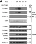
In Cell Death Dis on 20 January 2024 by Weinelt, N., Wächtershäuser, K. N., et al.
Fig.5.A

-
WB
-
Collected and cropped from Cell Death Dis by CiteAb, provided under a CC-BY license
Image 1 of 13
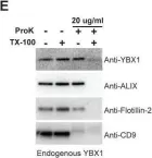
In Elife on 12 November 2021 by Liu, X. M., Ma, L., et al.
Fig.4.E

-
WB
-
Collected and cropped from Elife by CiteAb, provided under a CC-BY license
Image 1 of 13
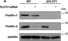
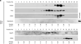
In Cell Mol Life Sci on 1 April 2021 by Brandel, A., Aigal, S., et al.
Fig.7.A

-
WB
-
Collected and cropped from Cell Mol Life Sci by CiteAb, provided under a CC-BY license
Image 1 of 13
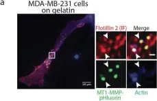
In Cancer Metastasis Rev on 1 June 2020 by Gauthier-Rouvière, C., Bodin, S., et al.
Fig.2.D

-
ICC-IF
-
Collected and cropped from Cancer Metastasis Rev by CiteAb, provided under a CC-BY license
Image 1 of 13
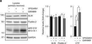
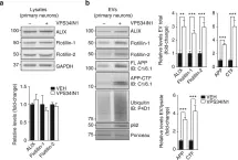
In Nat Commun on 18 January 2018 by Miranda, A. M., Lasiecka, Z. M., et al.
Fig.6.A

-
WB
-
Mus musculus (House mouse)
Collected and cropped from Nat Commun by CiteAb, provided under a CC-BY license
Image 1 of 13
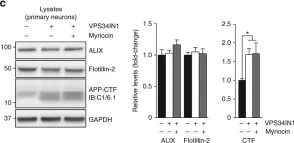
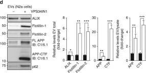
In Nat Commun on 18 January 2018 by Miranda, A. M., Lasiecka, Z. M., et al.
Fig.6.C

-
WB
-
Mus musculus (House mouse)
Collected and cropped from Nat Commun by CiteAb, provided under a CC-BY license
Image 1 of 13
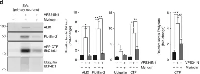
In Nat Commun on 18 January 2018 by Miranda, A. M., Lasiecka, Z. M., et al.
Fig.6.D

-
WB
-
Mus musculus (House mouse)
Collected and cropped from Nat Commun by CiteAb, provided under a CC-BY license
Image 1 of 13
In Nat Commun on 18 January 2018 by Miranda, A. M., Lasiecka, Z. M., et al.
Fig.5.A,B

-
WB
-
Mus musculus (House mouse)
Collected and cropped from Nat Commun by CiteAb, provided under a CC-BY license
Image 1 of 13
In Nat Commun on 18 January 2018 by Miranda, A. M., Lasiecka, Z. M., et al.
Fig.4.A,B

-
WB
-
Mus musculus (House mouse)
Collected and cropped from Nat Commun by CiteAb, provided under a CC-BY license
Image 1 of 13
In Nat Commun on 18 January 2018 by Miranda, A. M., Lasiecka, Z. M., et al.
Fig.4.D

-
WB
-
Mus musculus (House mouse)
Collected and cropped from Nat Commun by CiteAb, provided under a CC-BY license
Image 1 of 13
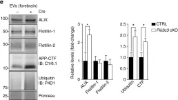
In Nat Commun on 18 January 2018 by Miranda, A. M., Lasiecka, Z. M., et al.
Fig.7.E

-
WB
-
Mus musculus (House mouse)
Collected and cropped from Nat Commun by CiteAb, provided under a CC-BY license
Image 1 of 13
In Sci Rep on 21 October 2016 by Chen, Q., Takada, R., et al.
Fig.2.A

-
WB
-
Canis lupus familiaris (Domestic dog)
Collected and cropped from Sci Rep by CiteAb, provided under a CC-BY license
Image 1 of 13
In Sci Rep on 21 October 2016 by Chen, Q., Takada, R., et al.
Fig.3.B

-
WB
-
Canis lupus familiaris (Domestic dog)
Collected and cropped from Sci Rep by CiteAb, provided under a CC-BY license
Image 1 of 13
