Cold acclimation increases insulin sensitivity, and some level of muscle contraction appears to be needed for provoking this effect. Here 15 men and (postmenopausal) women with overweight or obesity, the majority of whom had impaired glucose tolerance, were intermittently exposed to cold to induce 1 h of shivering per day over 10 days. We determined the effect of cold acclimation with shivering on overnight fasted oral glucose tolerance (primary outcome) and on skeletal muscle glucose transporter 4 translocation (secondary outcome). We find that cold acclimation with shivering improves oral glucose tolerance, fasting glucose, triglycerides, non-esterified fatty acid concentrations and blood pressure. Cold acclimation with shivering may thus represent an alternative lifestyle approach for the prevention and treatment of obesity-related metabolic disorders. ClinicalTrials.gov registration: NCT04516018 .
© 2024. The Author(s), under exclusive licence to Springer Nature Limited.
Product Citations: 61
Cold acclimation with shivering improves metabolic health in adults with overweight or obesity.
In Nature Metabolism on 1 December 2024 by Sellers, A. J., van Beek, S. M. M., et al.
-
Biochemistry and Molecular biology
-
Cell Biology
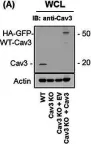
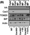
Cysteine post-translational modifications regulate protein interactions of caveolin-3.
In The FASEB Journal on 15 March 2024 by Ashford, F. B., Kuo, C. S., et al.
Caveolae are small flask-shaped invaginations of the surface membrane which are proposed to recruit and co-localize signaling molecules. The distinctive caveolar shape is achieved by the oligomeric structural protein caveolin, of which three isoforms exist. Aside from the finding that caveolin-3 is specifically expressed in muscle, functional differences between the caveolin isoforms have not been rigorously investigated. Caveolin-3 is relatively cysteine-rich compared to caveolins 1 and 2, so we investigated its cysteine post-translational modifications. We find that caveolin-3 is palmitoylated at 6 cysteines and becomes glutathiolated following redox stress. We map the caveolin-3 palmitoylation sites to a cluster of cysteines in its C terminal membrane domain, and the glutathiolation site to an N terminal cysteine close to the region of caveolin-3 proposed to engage in protein interactions. Glutathiolation abolishes caveolin-3 interaction with heterotrimeric G protein alpha subunits. Our results indicate that a caveolin-3 oligomer contains up to 66 palmitates, compared to up to 33 for caveolin-1. The additional palmitoylation sites in caveolin-3 therefore provide a mechanistic basis by which caveolae in smooth and striated muscle can possess unique phospholipid and protein cargoes. These unique adaptations of the muscle-specific caveolin isoform have important implications for caveolar assembly and signaling.
(c) 2024 The Authors. The FASEB Journal published by Wiley Periodicals LLC onbehalf of Federation of American Societies for Experimental Biology.
-
Biochemistry and Molecular biology
In FASEB BioAdvances on 1 March 2024 by Shi, J. & Wei, L.
In this study, we investigated the roles of ROCK1 in regulating structural and functional features of caveolae located at the cell membrane of cardiomyocytes, adipocytes, and mouse embryonic fibroblasts (MEFs) as well as related physiopathological effects. Caveolae are small bulb-shaped cell membrane invaginations, and their roles have been associated with disease conditions. One of the unique features of caveolae is that they are physically linked to the actin cytoskeleton that is well known to be regulated by RhoA/ROCKs pathway. In cardiomyocytes, we observed that ROCK1 deficiency is coincident with an increased caveolar density, clusters, and caveolar proteins including caveolin-1 and -3. In the mouse cardiomyopathy model with transgenic overexpressing Gαq in myocardium, we demonstrated the reduced caveolar density at cell membrane and reduced caveolar protein contents. Interestingly, coexisting ROCK1 deficiency in cardiomyocytes can rescue these defects and preserve caveolar compartmentalization of β-adrenergic signaling molecules including β1-adrenergic receptor and type V/VI adenylyl cyclase. In cardiomyocytes and adipocytes, we detected that ROCK1 deficiency increased insulin signaling with increased insulin receptor activation in caveolae. In MEFs, we identified that ROCK1 deficiency increased caveolar and total levels of caveolin-1 and cell membrane repair ability after mechanical or chemical disruptions. Together, these results demonstrate that ROCK1 can regulate caveolae plasticity and multiple functions including compartmentalization of signaling molecules and cell membrane repair following membrane disruption by mechanical force and oxidative damage. These findings provide possible molecular insights into the beneficial effects of ROCK1 deletion/inhibition in cardiomyocytes, adipocytes, and MEFs under certain diseased conditions.
© 2024 The Authors. FASEB BioAdvances published by Wiley Periodicals LLC on behalf of The Federation of American Societies for Experimental Biology.
-
WB
-
Mus musculus (House mouse)
Glutathione-dependent depalmitoylation of phospholemman by peroxiredoxin 6.
In Cell Reports on 27 February 2024 by Howie, J., Tulloch, L. B., et al.
Phospholemman (PLM) regulates the cardiac sodium pump: PLM phosphorylation activates the pump whereas PLM palmitoylation inhibits its activity. Here, we show that the anti-oxidant protein peroxiredoxin 6 (Prdx6) interacts with and depalmitoylates PLM in a glutathione-dependent manner. Glutathione loading cells acutely reduce PLM palmitoylation; glutathione depletion significantly increases PLM palmitoylation. Prdx6 silencing abolishes these effects, suggesting that PLM can be depalmitoylated by reduced Prdx6. In vitro, only recombinant Prdx6, among several peroxiredoxin isoforms tested, removes palmitic acid from recombinant palmitoylated PLM. The broad-spectrum depalmitoylase inhibitor palmostatin B prevents Prdx6-dependent PLM depalmitoylation in cells and in vitro. Our data suggest that Prdx6 is a thioesterase that can depalmitoylate proteins by nucleophilic attack via its reactive thiol, linking PLM palmitoylation and hence sodium pump activity to cellular glutathione status. We show that protein depalmitoylation can occur via a catalytic cysteine in which substrate specificity is determined by a protein-protein interaction.
Copyright © 2024 The Authors. Published by Elsevier Inc. All rights reserved.
In Antioxidants (Basel, Switzerland) on 31 January 2024 by Bartra, C., Yuan, Y., et al.
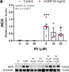
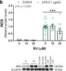
Resveratrol is a natural phenolic compound with known benefits against neurodegeneration. We analyzed in vitro the protective mechanisms of resveratrol against the proinflammatory monomeric C-reactive protein (mCRP). mCRP increases the risk of AD after stroke and we previously demonstrated that intracerebral mCRP induces AD-like dementia in mice. Here, we used BV2 microglia treated with mCRP for 24 h in the presence or absence of resveratrol. Cells and conditioned media were collected for analysis. Lipopolysaccharide (LPS) has also been implicated in AD progression and so LPS was used as a resveratrol-sensitive reference agent. mCRP at the concentration of 50 µg/mL activated the nitric oxide pathway and the NLRP3 inflammasome pathway. Furthermore, mCRP induced cyclooxygenase-2 and the release of proinflammatory cytokines. Resveratrol effectively inhibited these changes and increased the expression of the antioxidant enzyme genes Cat and Sod2. As central mechanisms of defense, resveratrol activated the hub genes Sirt1 and Nfe2l2 and inhibited the nuclear translocation of the signal transducer NF-ĸB. Proinflammatory changes induced by mCRP in primary mixed glial cultures were also protected by resveratrol. This work provides a mechanistic insight into the protective benefits of resveratrol in preventing the risk of AD induced by proinflammatory agents.
-
WB
-
Mus musculus (House mouse)
-
Immunology and Microbiology
-
Neuroscience
In Antioxidants (Basel) on 31 January 2024 by Bartra, C., Yuan, Y., et al.
Fig.3.A

-
WB
-
Collected and cropped from Antioxidants (Basel) by CiteAb, provided under a CC-BY license
Image 1 of 16
In Antioxidants (Basel) on 31 January 2024 by Bartra, C., Yuan, Y., et al.
Fig.3.B

-
WB
-
Collected and cropped from Antioxidants (Basel) by CiteAb, provided under a CC-BY license
Image 1 of 16
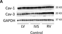
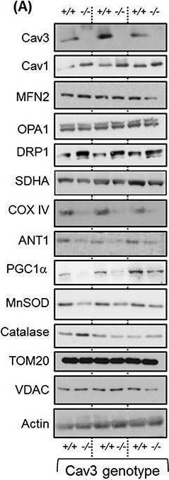
In J Cachexia Sarcopenia Muscle on 1 June 2020 by Shah, D. S., Nisr, R. B., et al.
Fig.7.A

-
WB
-
Collected and cropped from J Cachexia Sarcopenia Muscle by CiteAb, provided under a CC-BY license
Image 1 of 16
In J Cachexia Sarcopenia Muscle on 1 June 2020 by Shah, D. S., Nisr, R. B., et al.
Fig.7.B

-
WB
-
Collected and cropped from J Cachexia Sarcopenia Muscle by CiteAb, provided under a CC-BY license
Image 1 of 16
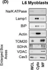
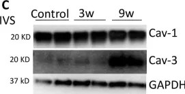
In J Cachexia Sarcopenia Muscle on 1 June 2020 by Shah, D. S., Nisr, R. B., et al.
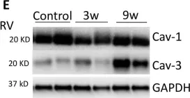
Fig.1.D

-
WB
-
Collected and cropped from J Cachexia Sarcopenia Muscle by CiteAb, provided under a CC-BY license
Image 1 of 16
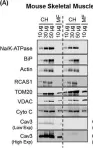
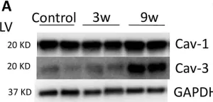
In J Cachexia Sarcopenia Muscle on 1 June 2020 by Shah, D. S., Nisr, R. B., et al.
Fig.1.A

-
WB
-
Collected and cropped from J Cachexia Sarcopenia Muscle by CiteAb, provided under a CC-BY license
Image 1 of 16
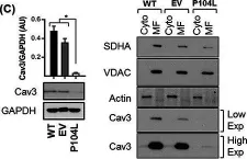
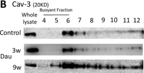
In J Cachexia Sarcopenia Muscle on 1 June 2020 by Shah, D. S., Nisr, R. B., et al.
Fig.3.C

-
WB
-
Collected and cropped from J Cachexia Sarcopenia Muscle by CiteAb, provided under a CC-BY license
Image 1 of 16
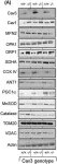
In J Cachexia Sarcopenia Muscle on 1 June 2020 by Shah, D. S., Nisr, R. B., et al.
Fig.4.A
-
WB
-
Collected and cropped from J Cachexia Sarcopenia Muscle by CiteAb, provided under a CC-BY license
Image 1 of 16
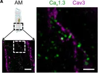
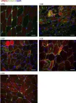
In Front Physiol on 24 October 2018 by Brandenburg, S., Pawlowitz, J., et al.
Fig.2.A

-
IHC-IF
-
Mus musculus (House mouse)
Collected and cropped from Front Physiol by CiteAb, provided under a CC-BY license
Image 1 of 16
In PLoS One on 13 May 2017 by Ichikawa, Y., Zemljic-Harpf, A. E., et al.
Fig.2.A

-
WB
-
Collected and cropped from PLoS One by CiteAb, provided under a CC-BY license
Image 1 of 16
In PLoS One on 13 May 2017 by Ichikawa, Y., Zemljic-Harpf, A. E., et al.
Fig.3.C

-
WB
-
Collected and cropped from PLoS One by CiteAb, provided under a CC-BY license
Image 1 of 16
In PLoS One on 13 May 2017 by Ichikawa, Y., Zemljic-Harpf, A. E., et al.
Fig.3.A

-
WB
-
Collected and cropped from PLoS One by CiteAb, provided under a CC-BY license
Image 1 of 16
In PLoS One on 13 May 2017 by Ichikawa, Y., Zemljic-Harpf, A. E., et al.
Fig.3.E

-
WB
-
Collected and cropped from PLoS One by CiteAb, provided under a CC-BY license
Image 1 of 16
In PLoS One on 13 May 2017 by Ichikawa, Y., Zemljic-Harpf, A. E., et al.
Fig.4.B

-
WB
-
Collected and cropped from PLoS One by CiteAb, provided under a CC-BY license
Image 1 of 16
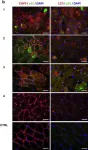
In Cell Death Dis on 19 January 2017 by Nascimbeni, A. C., Fanin, M., et al.
Fig.4.A

-
IHC-IF
-
Homo sapiens (Human)
Collected and cropped from Cell Death Dis by CiteAb, provided under a CC-BY license
Image 1 of 16
In Cell Death Dis on 19 January 2017 by Nascimbeni, A. C., Fanin, M., et al.
Fig.1.B

-
IHC-IF
-
Homo sapiens (Human)
Collected and cropped from Cell Death Dis by CiteAb, provided under a CC-BY license
Image 1 of 16