A critical step in cell morphogenesis is the extension of actin-dense pseudopods, controlled by actin-binding proteins (ABPs). While this process is well-understood on glass coverslips, it is less so in compliant three-dimensional environments. Here, we knocked out a series of ABPs in osteosarcoma cells and evaluated their effect on pseudopod extension on glass surfaces (2D) and in collagen gels (3D). Cells lacking the longest Arp3 gene variant, or with attenuated Arp2/3 activity, had the strongest reduction in pseudopod formation between 2D and 3D. This was largely due to reduced activity of the hybrid Arp2/3-vinculin complex, which was dispensable on glass. Our data suggests that concurrent formation of actin branches and nascent adhesions, supported by Arp2/3-vinculin interactions, is essential to form mechanically stable links between fibrous extracellular matrix and actin in 3D. This highlights how experiments on stiff, planar substrates may conceal actin architectural features that are essential for morphogenesis in 3D.
© 2025 The Authors. Published by Elsevier Inc.
Product Citations: 15
In IScience on 20 June 2025 by Isogai, T., Dean, K. M., et al.
Phosphoserine-86-HSPB1 (pS86-HSPB1) is cytoplasmic and highly induced in rat myometrium at labour.
In Histochemistry and Cell Biology on 1 February 2023 by Miskiewicz, E. I., Olaloku, A., et al.
Uterine myocytes during pregnancy proceed through a series of adaptations and collectively transform into a powerfully contractile tissue by term. Previous work has indicated that members of the heat shock protein (HSP) B family of stress proteins are associated with the process of adaptation and transformation. Utilizing immunoblot analyses, widefield epifluorescence and total internal reflection (TIRF) microscopy, this study investigated the temporal and spatial detection of HSPB1 phosphorylated on serine-86 (pS86-HSPB1) in rat myometrium during pregnancy, the role of uterine distension in regulation of pS86-HSPB1, and the comparative localization with pS15-HSPB1 in rat myometrial tissue as well as in an immortalized human myometrial cell line. Immunoblot detection of pS86-HSPB1 was significantly elevated during late pregnancy and labour. In particular, pS86-HSPB1 was significantly increased at day (d)22 and d23 (labour) compared with all other timepoints assessed. Localization of pS86-HSPB1 in myometrium became prominent at d22 and d23 with cytoplasmic detection around myometrial cell nuclei. Furthermore, pS86-HSPB1 detection was found to be significantly elevated in the gravid rat uterine myometrium compared with the non-gravid tissue at d19 and d23. Both widefield epifluorescence and TIRF microscopy examination of human myometrial cells demonstrated that pS15-HSPB1 was prominently localized to focal adhesions, while pS82-HSPB1 (homologous to rodent pS86-HSPB1) was primarily located in the cell cytoplasm. Our data demonstrate that levels of phosphorylated HSPB1 increase just prior to and during labour, and that uterine distension is a stress-inducing signal for HSPB1 phosphorylation. The exact roles of these phosphorylated forms in myometrial cells remain to be determined.
© 2022. The Author(s).
-
Cell Biology
Sensing Actin Dynamics through Adherens Junctions.
In Cell Reports on 25 February 2020 by Indra, I., Troyanovsky, R. B., et al.
We study punctate adherens junctions (pAJs) to determine how short-lived cadherin clusters and relatively stable actin bundles interact despite differences in dynamics. We show that pAJ-linked bundles consist of two distinct regions-the bundle stalk (AJ-BS) and a tip (AJ-BT) positioned between cadherin clusters and the stalk. The tip differs from the stalk in a number of ways: it is devoid of the actin-bundling protein calponin, and exhibits a much faster F-actin turnover rate. While F-actin in the stalk displays centripetal movement, the F-actin in the tip is immobile. The F-actin turnover in both the tip and stalk is dependent on cadherin cluster stability, which in turn is regulated by F-actin. The close bidirectional coupling between the stability of cadherin and associated F-actin shows how pAJs, and perhaps other AJs, allow cells to sense and coordinate the dynamics of the actin cytoskeleton in neighboring cells-a mechanism we term "dynasensing."
Copyright © 2020 The Authors. Published by Elsevier Inc. All rights reserved.
-
Homo sapiens (Human)
-
Cell Biology
In Theranostics on 4 October 2018 by Liu, Z., Wang, Y., et al.
Rational: Patients with hepatocellular carcinoma (HCC) have a poor prognosis mostly due to intrahepatic as well as distal metastasis. Vasodilator-stimulated phosphoprotein (VASP), a regulator of actin cytoskeleton and cell migration, is overexpressed in HCC and correlated with its malignant features and poor prognosis. Very little is known about its function in HCC. Methods: qRT-PCR, Western blot and IHC were used to detect the VASP expression in tissues and cells. Transwell and wound healing assays were used to measure the migration and invasion of HCC cells. Immunoblotting and immunofluorescence were used for detection of epithelial-to-mesenchymal transition (EMT) progression in HCC cells. A lung metastasis mouse model was used to evaluate metastasis of HCC in vivo. The putative targets of miR-204 were disclosed by public databases and a dual-luciferase reporter assay. IP was used to show the interaction between VASP and CRKL. ChIP was used to analyze the binding of HIF-1α to VASP promoter region. Results: Our data involving both gain- and loss-of-function studies revealed that VASP activated AKT and ERK signaling and promoted HCC migration and invasion in vitro and in vivo by altering the EMT phenotype and expression of MMPs. We investigated the positive correlation between VASP and an adapter protein, CRKL. VASP dynamically co-localized at the SH3N domain of CRKL and mediated its function. Mechanistically, VASP overexpression at the transcriptional level was mediated by HIF-1α through direct binding to two hypoxia response elements (HRE) in the VASP promoter region. Furthermore, we identified hypoxia-induced down-regulation of miR-204, which functioned as the regulator of VASP overexpression at the post-transcriptional level. Also, hypoxia-activated p-Smad3 dependent TGF-β signaling indirectly promoted VASP expression. Conclusion: A variety of hypoxia-induced molecular mechanisms contributed to the upregulation of VASP at transcriptional and post-transcriptional levels. These mechanisms involved CRKL, HIF-1α, miR-204, and TGF-β activating the AKT and ERK signaling to promote EMT and expression of MMPs. Taken together, our results defined VASP as an oncogene of HCC pathogenesis and metastasis with the potential to serve as a prognostic biomarker.
-
IP
-
Cancer Research
In NPJ Precision Oncology on 7 June 2018 by Xiang, X., Wang, Y., et al.
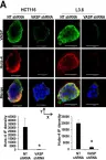
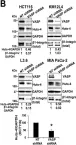
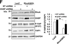
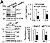
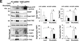
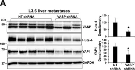
Extracellular matrix (ECM)-induced β1-integrin-FAK signaling promotes cell attachment, survival, and migration of cancer cells in a distant organ so as to enable cancer metastasis. However, mechanisms governing activation of the β1-integrin-FAK signaling remain incompletely understood. Here, we report that vasodilator-stimulated phosphoprotein (VASP), an actin binding protein, is required for ECM-mediated β1-integrin-FAK-YAP1/TAZ signaling in gastrointestinal (GI) cancer cells and their liver metastasis. In patient-derived samples, VASP is upregulated in 53 of 63 colorectal cancers and 43 of 53 pancreatic ductal adenocarcinomas and high VASP levels correlate with liver metastasis and reduced patient survival. In a Matrigel-based 3-dimensional (3D) culture model, short hairpin RNA (shRNA)-mediated VASP knockdown in colorectal cancer cells (KM12L4, HCT116, and HT29) and pancreatic cancer cells (L3.6 and MIA PaCa-1) suppresses the growth of 3D cancer spheroids. Mechanistic studies reveal that VASP knockdown suppresses FAK phosphorylation and YAP1/TAZ protein levels, but not Akt or Erk-related pathways and that YAP1/TAZ proteins are enhanced by the β1-integrin-FAK signaling. Additionally, VASP regulates the β1-integrin-FAK-YAP1/TAZ signaling by at least two mechanisms: (1) promoting ECM-mediated β1-integrin activation and (2) regulating YAP1/TAZ dephosphorylation at downstream of RhoA to enhance the stability of YAP1/TAZ proteins. In agreement with these, preclinical studies with two experimental liver metastasis mouse models demonstrate that VASP knockdown suppresses GI cancer liver metastasis, β1-integrin activation, and YAP1/TAZ levels of metastatic cancer cells. Together, our data support VASP as a treatment target for liver metastasis of colorectal and pancreatic cancers.
-
ICC-IF
-
WB
-
Cancer Research
In NPJ Precis Oncol on 7 June 2018 by Xiang, X., Wang, Y., et al.
Fig.3.A

-
ICC-IF
-
Collected and cropped from NPJ Precis Oncol by CiteAb, provided under a CC-BY license
Image 1 of 6
In NPJ Precis Oncol on 7 June 2018 by Xiang, X., Wang, Y., et al.
Fig.3.B

-
WB
-
Collected and cropped from NPJ Precis Oncol by CiteAb, provided under a CC-BY license
Image 1 of 6
In NPJ Precis Oncol on 7 June 2018 by Xiang, X., Wang, Y., et al.
Fig.4.F

-
WB
-
Collected and cropped from NPJ Precis Oncol by CiteAb, provided under a CC-BY license
Image 1 of 6
In NPJ Precis Oncol on 7 June 2018 by Xiang, X., Wang, Y., et al.
Fig.4.A

-
WB
-
Collected and cropped from NPJ Precis Oncol by CiteAb, provided under a CC-BY license
Image 1 of 6
In NPJ Precis Oncol on 7 June 2018 by Xiang, X., Wang, Y., et al.
Fig.4.E

-
WB
-
Collected and cropped from NPJ Precis Oncol by CiteAb, provided under a CC-BY license
Image 1 of 6
In NPJ Precis Oncol on 7 June 2018 by Xiang, X., Wang, Y., et al.
Fig.6.A

-
WB
-
Collected and cropped from NPJ Precis Oncol by CiteAb, provided under a CC-BY license
Image 1 of 6