Talin-1 (TLN1) is best known to activate integrin receptors and transmit mechanical stimuli to the actin cytoskeleton at focal adhesions. However, the localization of TLN1 is not restricted to focal adhesions. By utilizing both subcellular fractionations and confocal microscopy analyses, we show that TLN1 localizes to the nucleus in several human cell lines, where it is tightly associated with the chromatin. Importantly, small interfering RNA (siRNA)-mediated depletion of endogenous TLN1 triggers extensive changes in the gene expression profile of human breast epithelial cells. To determine the functional impact of nuclear TLN1, we expressed a TLN1 fusion protein containing a nuclear localization signal. Our findings revealed that the accumulation of nuclear TLN1 alters the expression of a subset of genes and impairs the formation of cell-cell clusters. This study introduces an additional perspective on the canonical view of TLN1 subcellular localization and function.
© 2025 The Author(s).
Product Citations: 68
Nuclear talin-1 provides a bridge between cell adhesion and gene expression.
In IScience on 21 February 2025 by Da Silva, A. J., Hästbacka, H. S. E., et al.
In Antioxidants (Basel, Switzerland) on 20 January 2025 by Xu, H., Lan, S., et al.
Andrographis paniculata is mainly used to treat skin inflammations, wounds, and infections. In this study, Andrographis Herba, the aerial part of the plant, was proven to increase the viability of UVB-damaged HaCat cells and reduce reactive oxygen species levels. The chemical composition of Andrographis Herba extract (AHE) was analyzed using UPLC-Q-TOF-MS, and diterpene lactones were identified as its primary constituents. Then, the fraction of diterpene lactones was prepared and exhibited similar effects to AHE. AHE, its diterpene lactones component, and its representative constituent andrographolide all decreased the expression of IL-1β, IL-6, and CDKN1A. Furthermore, the protective effects of AHE and its active ingredients on UVB-damaged epidermal stem cells were investigated. Notably, the combined treatment with andrographolide and collagen XVII enhanced the viability of UVB-damaged epidermal stem cells, increased the expression of stemness markers integrin β1 and p63, and decreased the expression of the differentiation marker keratin 10. This combination demonstrated significant synergy in maintaining skin homeostasis, which provides evidences for the development of skin-protective products.
In Animals : An Open Access Journal From MDPI on 26 November 2024 by Jiang, E., Chen, X., et al.
In livestock production, oxidative stress (OS) is ubiquitous, reducing animal productivity and product quality. Hence, investigating the mechanisms of oxidative stress in livestock and inhibiting oxidative stress-induced damage is crucial. Curcumin, a plant-derived bioactive compound, exhibits antioxidant and anti-apoptotic properties. Adipose-derived stem cells (ADSCs) from animal adipose tissue are easily accessible and possess multilineage differentiation potential. Therefore, this work utilized bovine ADSCs to establish an oxidative stress model and investigated the effects of curcumin on oxidative stress and apoptosis. Firstly, bovine ADSCs were isolated and cultured from fetal calf subcutaneous adipose tissue. Their surface markers were identified by immunofluorescence, confirming the expression of CD29, CD44, CD73, CD90, CD105 and Vimentin, but not CD34, indicative of mesenchymal stem/progenitor cell characteristics. Secondly, to explore the effects of curcumin on oxidative damage and apoptosis in bovine ADSCs, an oxidative stress model was induced using H2O2. CCK-8 assays showed significantly reduced cell viability and SOD activity, along with increased malondialdehyde (MDA) and reactive oxygen species (ROS) levels, indicating successful modeling. RT-qPCR further confirmed that 500 μM of H2O2 treatment for 24 h promoted apoptosis. Herein, CCK-8 assays indicated a significant reduction in cell viability at >8 μM of curcumin. Thirdly, using 4 μM and 8 μM of curcumin for pre-protection, 8 μM maintained SOD activity, reduced MDA and ROS, inhibited apoptosis-related gene changes (Bcl-2, Bax, Caspase-3), and suppressed apoptosis according to a TUNEL assay. Fourthly, curcumin's autophagy-inducing potential was hypothesized, which was confirmed by increased LC3-II and decreased P62 expression upon co-treatment with 3-MA. 3-MA inhibited curcumin's antioxidant and anti-apoptotic effects, suggesting that curcumin's antioxidant and anti-apoptotic roles may involve autophagy induction. In conclusion, bovine ADSCs are abundant, easily accessible, and multipotent, making them suitable for in vitro expansion. Curcumin alleviated H2O2-induced oxidative stress in bovine ADSCs, with curcumin also inhibiting apoptosis, likely through autophagy induction. This study validates the protective role of curcumin in bovine ADSCs, with potential applications in livestock production.
-
Stem Cells and Developmental Biology
-
Veterinary Research
Preprint on BioRxiv : the Preprint Server for Biology on 21 November 2024 by Pasquier, N., Isomursu, A., et al.
Mucinous colorectal carcinoma (MUC CRC) dissemination into the tumor stroma and metastasis to multiple organs, including the peritoneum, is associated with poor prognosis. Disseminating MUC CRCs exhibit either a conventional ‘apical-in’ or an inverted ‘apical-out’ polarity phenotype that influence patient outcome. Identifying the mechanisms controlling MUC CRC polarity is critical to understand disease progression. Here, we analyze patient-derived MUC CRC xenografts, with apical-in or apical-out polarity, ex vivo or within collagen gels to mimic the peritumoral stroma. Single-cell analyses reveal α2β1-integrin as a key collagen-binding receptor in these models. Collagen–α2β1-integrin interaction activates Src and ERK/MAPK signaling and upregulates the expression of SorLA, an endosomal sorting receptor. SorLA supports apical-in polarity and carcinoma-stroma interactions by promoting integrin recycling to the plasma membrane and HER2/HER3 expression through a positive feedback mechanism. Accordingly, we observe positive correlation between HER2, HER3 and SorLA in patient samples with the highest HER2 expression in apical-in-presenting tissues. Treatment of tumor spheres with clinically relevant HER2/HER3-targeting antibodies reverts sphere polarity and impedes collagen remodeling and adhesion to mouse peritoneum. This SorLA—integrin—HER2/HER3 signaling axis may represent a basis for MUC CRC-patient stratification and shed light on other carcinomas with similar apical-out phenotypes.
-
Cancer Research
Diverse Fgfr1 signaling pathways and endocytic trafficking regulate mesoderm development.
In Genes and Development on 25 June 2024 by Clark, J. F. & Soriano, P.
The fibroblast growth factor (FGF) pathway is a conserved signaling pathway required for embryonic development. Activated FGF receptor 1 (FGFR1) drives multiple intracellular signaling cascade pathways, including ERK/MAPK and PI3K/AKT, collectively termed canonical signaling. However, unlike Fgfr1-null embryos, embryos containing hypomorphic mutations in Fgfr1 lacking the ability to activate canonical downstream signals are still able to develop to birth but exhibit severe defects in all mesodermal-derived tissues. The introduction of an additional signaling mutation further reduces the activity of Fgfr1, leading to earlier lethality, reduced somitogenesis, and more severe changes in transcriptional outputs. Genes involved in migration, ECM interaction, and phosphoinositol signaling were significantly downregulated, proteomic analysis identified changes in interactions with endocytic pathway components, and cells expressing mutant receptors show changes in endocytic trafficking. Together, we identified processes regulating early mesoderm development by mechanisms involving both canonical and noncanonical Fgfr1 pathways, including direct interaction with cell adhesion components and endocytic regulation.
© 2024 Clark and Soriano; Published by Cold Spring Harbor Laboratory Press.
-
Mus musculus (House mouse)
-
Stem Cells and Developmental Biology
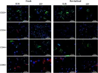
In Stem Cell Rev Rep on 1 April 2016 by Duan, W. & Lopez, M. J.
Fig.7.A

-
ICC
-
Canis lupus familiaris (Domestic dog)
Collected and cropped from Stem Cell Rev Rep by CiteAb, provided under a CC-BY license
Image 1 of 2
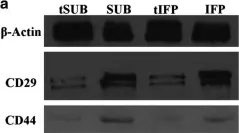
In Stem Cell Rev Rep on 1 April 2016 by Duan, W. & Lopez, M. J.
Fig.9.A

-
WB
-
Canis lupus familiaris (Domestic dog)
Collected and cropped from Stem Cell Rev Rep by CiteAb, provided under a CC-BY license
Image 1 of 2