Defective renal excretion and increased production of uric acid engender hyperuricemia that predisposes to gout. However, molecular mechanisms underlying defective uric acid excretion remain largely unknown. Here, we report a rare genetic variant of gout-unprecedented NUMB gene within a hereditary human gout family, which was identified by an unbiased genome-wide sequencing approach. This dysfunctional missense variant within the conserved region of the NUMB gene (NUMBR630H) underwent intracellular redistribution and degradation through an autophagy-dependent mechanism. Mechanistically, we identified the uric acid transporter, ATP Binding Cassette Subfamily G Member 2 (ABCG2), as a novel NUMB-binding protein through its intracellular YxNxxF motif. In polarized renal tubular epithelial cells (RTECs), NUMB promoted ABCG2 trafficking towards the apical plasma membrane. Genetic loss-of-function of NUMB resulted in redistribution of ABCG2 in the basolateral domain and ultimately defective excretion of uric acid. To recapitulate the clinical situation in human gout patients, we generated a NUMBR630H knock-in mouse strain, which showed marked increases of serum urate and decreased uric acid excretion. The NUMBR630H knock-in mice exhibited clinically relevant hyperuricemia. In summary, we have uncovered a novel NUMB-mediated mechanism of uric acid excretion and a functional missense variant of NUMB in humans, which causes hyperuricemia and gout.
© 2024. The Author(s).
Product Citations: 33
NUMB dysfunction defines a novel mechanism underlying hyperuricemia and gout.
In Cell Discovery on 22 October 2024 by Chi, J., Chen, Y., et al.
Identification of the growth cone as a probe and driver of neuronal migration in the injured brain.
In Nature Communications on 9 March 2024 by Nakajima, C., Sawada, M., et al.
Axonal growth cones mediate axonal guidance and growth regulation. We show that migrating neurons in mice possess a growth cone at the tip of their leading process, similar to that of axons, in terms of the cytoskeletal dynamics and functional responsivity through protein tyrosine phosphatase receptor type sigma (PTPσ). Migrating-neuron growth cones respond to chondroitin sulfate (CS) through PTPσ and collapse, which leads to inhibition of neuronal migration. In the presence of CS, the growth cones can revert to their extended morphology when their leading filopodia interact with heparan sulfate (HS), thus re-enabling neuronal migration. Implantation of an HS-containing biomaterial in the CS-rich injured cortex promotes the extension of the growth cone and improve the migration and regeneration of neurons, thereby enabling functional recovery. Thus, the growth cone of migrating neurons is responsive to extracellular environments and acts as a primary regulator of neuronal migration.
© 2024. The Author(s).
-
ICC
-
Mus musculus (House mouse)
In Communications Biology on 24 February 2024 by Kulicke, C. A., Swarbrick, G., et al.
MR1-restricted T cells have been implicated in microbial infections, sterile inflammation, wound healing and cancer. Similar to other antigen presentation molecules, evidence supports multiple, complementary MR1 antigen presentation pathways. To investigate ligand exchange pathways for MR1, we used MR1 monomers and tetramers loaded with 5-(2-oxopropylideneamino)-6-d-ribitylaminouracil (5-OP-RU) to deliver the antigen. Using MR1-deficient cells reconstituted with wild-type MR1 or MR1 molecules that cannot bind 5-OP-RU, we show that presentation of monomer-delivered 5-OP-RU is dependent on cellular MR1 and requires the transfer of ligand from the soluble molecule onto MR1 expressed by the antigen presenting cell. This mode of antigen delivery strengthens the evidence for post-ER ligand exchange pathways for MR1, which could represent an important avenue by which MR1 acquires antigens derived from endocytosed pathogens.
© 2024. This is a U.S. Government work and not under copyright protection in the US; foreign copyright protection may apply.
Synaptotagmin-11 Inhibits Synaptic Vesicle Endocytosis via Endophilin A1.
In The Journal of Neuroscience on 6 September 2023 by Wang, Y., Zhu, Y., et al.
Synaptic vesicle (SV) endocytosis is a critical and well-regulated process for the maintenance of neurotransmission. We previously reported that synaptotagmin-11 (Syt11), an essential non-Ca2+-binding Syt associated with brain diseases, inhibits neuronal endocytosis (Wang et al., 2016). Here, we found that Syt11 deficiency caused accelerated SV endocytosis and vesicle recycling under sustained stimulation and led to the abnormal membrane partition of synaptic proteins in mouse hippocampal boutons of either sex. Furthermore, our study revealed that Syt11 has direct but Ca2+-independent binding with endophilin A1 (EndoA1), a membrane curvature sensor and endocytic protein recruiter, with high affinity. EndoA1-knockdown significantly reversed Syt11-KO phenotype, identifying EndoA1 as a main inhibitory target of Syt11 during SV endocytosis. The N-terminus of EndoA1 and the C2B domain of Syt11 were responsible for this interaction. A peptide (amino acids 314-336) derived from the Syt11 C2B efficiently blocked Syt11-EndoA1 binding both in vitro and in vivo Application of this peptide inhibited SV endocytosis in WT hippocampal neurons but not in EndoA1-knockdown neurons. Moreover, intracellular application of this peptide in mouse calyx of Held terminals of either sex effectively hampered both fast and slow SV endocytosis at physiological temperature. We thus propose that Syt11 ensures the precision of protein retrieval during SV endocytosis by inhibiting EndoA1 function at neuronal terminals.SIGNIFICANCE STATEMENT Endocytosis is a key stage of synaptic vesicle (SV) recycling. SV endocytosis retrieves vesicular membrane and protein components precisely to support sustained neurotransmission. However, the molecular mechanisms underlying the regulation of SV endocytosis remain elusive. Here, we reported that Syt11-KO accelerated SV endocytosis and impaired membrane partition of synaptic proteins. EndoA1 was identified as a main inhibitory target of Syt11 during SV endocytosis. Our study reveals a novel inhibitory mechanism of SV endocytosis in preventing hyperactivation of endocytosis, potentially safeguarding the recycling of synaptic proteins during sustained neurotransmission.
Copyright © 2023 the authors.
-
Neuroscience
In The Journal of Membrane Biology on 1 October 2022 by Andhare, D. S., Khurana, H., et al.
Discovery-based proteomics workflows that identify novel interactors rely on immunoprecipitations or pull-downs with genetically tagged bait proteins immobilized on appropriate matrices. But strategies to analyse protein interactions on a diffusible-membrane surface combined with the practical ease of pull-downs remain unavailable. Such strategies are important to analyse protein complexes that mature in composition and stability because of diffusion-based encounter between participant proteins. Here, we describe a generic pull-down strategy to analyse such complexes using chelating lipid-containing supported bilayers formed on silica beads. These templates can display desired His-tagged bait proteins on a diffusible-membrane surface. Using clathrin-mediated endocytosis as a paradigm, we find that the clathrin-binding adaptor protein epsin1 displayed as bait on these templates pulls down significantly higher amounts of clathrin from brain lysates than when immobilized on conventional matrices. Together, our results establish the potential of such templates as superior matrices for analysing protein-protein interactions and resultant complexes formed on membrane surfaces.
© 2022. The Author(s), under exclusive licence to Springer Science+Business Media, LLC, part of Springer Nature.
-
WB
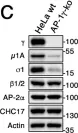
In Life Sci Alliance on 1 March 2022 by Buser, D. P., Bader, G., et al.
Fig.3.C

-
WB
-
Homo sapiens (Human)
Collected and cropped from Life Sci Alliance by CiteAb, provided under a CC-BY license
Image 1 of 1