Platelet-producing megakaryocytes (MKs) primarily reside in the bone marrow, where they duplicate their DNA content with each cell cycle resulting in polyploid cells with an intricate demarcation membrane system. While key elements of the cytoskeletal reorganizations during proplatelet formation have been identified, what initiates the release of platelets into vessel sinusoids remains largely elusive. Using a cell cycle indicator, we observed a unique phenomenon, during which amplified centrosomes in MKs underwent clustering following mitosis, closely followed by proplatelet formation, which exclusively occurred in G1 of interphase. Forced cell cycle arrest in G1 increased proplatelet formation not only in vitro but also in vivo following short-term starvation of mice. We identified that inhibition of the centrosomal protein kinesin family member C1 (KIFC1) impaired clustering and subsequent proplatelet formation, while KIFC1-deficient mice exhibited reduced platelet counts. In summary, we identified KIFC1- and cell cycle-mediated centrosome clustering as an important initiator of proplatelet formation from MKs.
Product Citations: 85
Cell cycle-dependent centrosome clustering precedes proplatelet formation.
In Science Advances on 21 June 2024 by Becker, I. C., Wilkie, A. R., et al.
-
Cell Biology
Molecular basis promoting centriole triplet microtubule assembly.
In Nature Communications on 22 March 2024 by Takeda, Y., Harada, T., et al.
The triplet microtubule, a core structure of centrioles crucial for the organization of centrosomes, cilia, and flagella, consists of unclosed incomplete microtubules. The mechanisms of its assembly represent a fundamental open question in biology. Here, we discover that the ciliopathy protein HYLS1 and the β-tubulin isotype TUBB promote centriole triplet microtubule assembly. HYLS1 or a C-terminal tail truncated version of TUBB generates tubulin-based superstructures composed of centriole-like incomplete microtubule chains when overexpressed in human cells. AlphaFold-based structural models and mutagenesis analyses further suggest that the ciliopathy-related residue D211 of HYLS1 physically traps the wobbling C-terminal tail of TUBB, thereby suppressing its inhibitory role in the initiation of the incomplete microtubule assembly. Overall, our findings provide molecular insights into the biogenesis of atypical microtubule architectures conserved for over a billion years.
© 2024. The Author(s).
-
Homo sapiens (Human)
In IScience on 15 March 2024 by Islam, A., Manjarrez-González, J. C., et al.
Chromosomal instability (CIN) is a hallmark of cancers, and CIN-promoting mutations are not fully understood. Here, we report 141 chromosomal instability aiding variant (CIVa) candidates by assessing the prevalence of loss-of-function (LoF) variants in 135 chromosome segregation genes from over 150,000 humans. Unexpectedly, we observe both heterozygous and homozygous CIVa in Astrin and SKA3, two evolutionarily conserved kinetochore and microtubule-associated proteins essential for chromosome segregation. To stratify harmful versus harmless variants, we combine live-cell microscopy and controlled protein expression. We find the naturally occurring Astrin p.Q1012∗ variant is harmful as it fails to localize normally and induces chromosome misalignment and missegregation, in a dominant negative manner. In contrast, the Astrin p.L7Qfs∗21 variant generates a shorter isoform that localizes and functions normally, and the SKA3 p.Q70Kfs∗7 variant allows wild-type SKA complex localisation and function, revealing distinct resilience mechanisms that render these variants harmless. Thus, we present a scalable framework to predict and stratify naturally occurring CIVa, and provide insight into resilience mechanisms that compensate for naturally occurring CIVa.
© 2024 The Author(s).
-
Homo sapiens (Human)
-
Genetics
A farnesyl-dependent structural role for CENP-E in expansion of the fibrous corona.
In The Journal of Cell Biology on 1 January 2024 by Wu, J., Raas, M. W. D., et al.
Correct chromosome segregation during cell division depends on proper connections between spindle microtubules and kinetochores. During prometaphase, kinetochores are temporarily covered with a dense protein meshwork known as the fibrous corona. Formed by oligomerization of ROD/ZW10/ZWILCH-SPINDLY (RZZ-S) complexes, the fibrous corona promotes spindle assembly, chromosome orientation, and spindle checkpoint signaling. The molecular requirements for formation of the fibrous corona are not fully understood. Here, we show that the fibrous corona depends on the mitotic kinesin CENP-E and that poorly expanded fibrous coronas after CENP-E depletion are functionally compromised. This previously unrecognized role for CENP-E does not require its motor activity but instead is driven by farnesyl modification of its C-terminal kinetochore- and microtubule-binding domain. We show that in cells, CENP-E binds Spindly and recruits RZZ-S complexes to ectopic locations in a farnesyl-dependent manner. CENP-E is recruited to kinetochores following RZZ-S, and-while not required for RZZ-S oligomerization per se-promotes subsequent fibrous corona expansion. Our comparative genomics analyses suggest that the farnesylation motif in CENP-E orthologs emerged alongside the full RZZ-S module in an ancestral lineage close to the fungi-animal split (Obazoa), revealing potential conservation of the mechanisms for fibrous corona formation. Our results show that proper spindle assembly has a potentially conserved non-motor contribution from the kinesin CENP-E through stabilization of the fibrous corona meshwork during its formation.
© 2023 Wu et al.
-
Homo sapiens (Human)
-
Cell Biology
PLK1 inhibition dampens NLRP3 inflammasome-elicited response in inflammatory disease models.
In The Journal of Clinical Investigation on 1 November 2023 by Baldrighi, M., Doreth, C., et al.
Unabated activation of the NLR family pyrin domain-containing 3 (NLRP3) inflammasome is linked with the pathogenesis of various inflammatory disorders. Polo-like kinase 1 (PLK1) has been widely studied for its role in mitosis. Here, using both pharmacological and genetic approaches, we demonstrate that PLK1 promoted NLRP3 inflammasome activation at cell interphase. Using an unbiased proximity-dependent biotin identification (Bio-ID) screen for the PLK1 interactome in macrophages, we show an enhanced proximal association of NLRP3 with PLK1 upon NLRP3 inflammasome activation. We further confirmed the interaction between PLK1 and NLRP3 and identified the interacting domains. Mechanistically, we show that PLK1 orchestrated the microtubule-organizing center (MTOC) structure and NLRP3 subcellular positioning upon inflammasome activation. Treatment with a selective PLK1 kinase inhibitor suppressed IL-1β production in in vivo inflammatory models, including LPS-induced endotoxemia and monosodium urate-induced peritonitis in mice. Our results uncover a role of PLK1 in regulating NLRP3 inflammasome activation during interphase and identify pharmacological inhibition of PLK1 as a potential therapeutic strategy for inflammatory diseases with excessive NLRP3 inflammasome activation.
-
Immunology and Microbiology
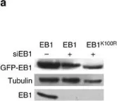
In Nat Commun on 19 September 2016 by Courtheoux, T., Enchev, R. I., et al.
Fig.4.A

-
WB
-
Homo sapiens (Human)
Collected and cropped from Nat Commun by CiteAb, provided under a CC-BY license
Image 1 of 9
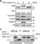
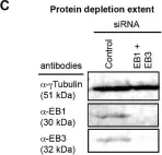
In Nat Commun on 31 March 2016 by Iimori, M., Watanabe, S., et al.
Fig.1.B,C

-
WB
-
Homo sapiens (Human)
Collected and cropped from Nat Commun by CiteAb, provided under a CC-BY license
Image 1 of 9
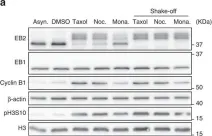
In Nat Commun on 31 March 2016 by Iimori, M., Watanabe, S., et al.
Fig.2.A

-
WB
-
Homo sapiens (Human)
Collected and cropped from Nat Commun by CiteAb, provided under a CC-BY license
Image 1 of 9
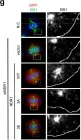
In Nat Commun on 21 October 2015 by Bizet, A. A., Becker-Heck, A., et al.
Fig.5.F

-
WB
-
Homo sapiens (Human)
Collected and cropped from Nat Commun by CiteAb, provided under a CC-BY license
Image 1 of 9
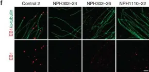
In Sci Rep on 9 June 2015 by Yan, M., Chu, L., et al.
Fig.5.G

-
ICC-IF
-
Homo sapiens (Human)
Collected and cropped from Sci Rep by CiteAb, provided under a CC-BY license
Image 1 of 9
In Biol Open on 16 January 2015 by Tamura, N., Simon, J. E., et al.
Fig.5.C

-
WB
-
Homo sapiens (Human)
Collected and cropped from Biol Open by CiteAb, provided under a CC-BY license
Image 1 of 9
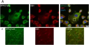
In PLoS One on 23 September 2014 by Poliakova, K., Adebola, A., et al.
Fig.4.A

-
ICC-IF
-
Collected and cropped from PLoS One by CiteAb, provided under a CC-BY license
Image 1 of 9
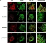
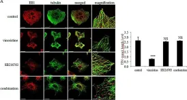
In Oncotarget on 30 May 2014 by Le Grand, M., Rovini, A., et al.
Fig.2.A

-
ICC-IF
-
Collected and cropped from Oncotarget by CiteAb, provided under a CC-BY license
Image 1 of 9
In Oncotarget on 30 May 2014 by Le Grand, M., Rovini, A., et al.
Fig.6.A

-
ICC-IF
-
Collected and cropped from Oncotarget by CiteAb, provided under a CC-BY license
Image 1 of 9