Idiopathic pulmonary fibrosis (IPF) is a chronic and refractory interstitial lung disease. Although there are two approved drugs for IPF, they were not able to completely cure the disease. Therefore, the development of new drugs is required for the effective treatment of IPF. In this study, we investigated the effect of theophylline, which has long been used for the treatment of asthma, on pulmonary fibrosis. The administration of theophylline attenuated the fibrotic changes of lung tissues and improved mechanical pulmonary functions in bleomycin (BLM)-induced pulmonary fibrosis. Theophylline treatment suppressed IL-17 production through inhibiting cytokines controlling Th17 differentiation; TGF-β, IL-6, IL-1β, and IL-23. The inhibition of IL-6 and IL-1β by theophylline is mediated by suppressing BLM-induced ROS production and NF-κB activation in epithelial cells. We further demonstrated that theophylline inhibited TGF-β-induced epithelial-to-mesenchymal transition in epithelial cells through suppressing the phosphorylation of Smad2/3 and AKT. The inhibitory effects of theophylline on the phosphorylation of Smad2/3 and AKT were recapitulated in BLM-treated lung tissues. Taken together, these results demonstrated that theophylline prevents pulmonary fibrosis by inhibiting Th17 differentiation and TGF-β signaling.
Product Citations: 26
Theophylline Attenuates BLM-Induced Pulmonary Fibrosis by Inhibiting Th17 Differentiation.
In International Journal of Molecular Sciences on 5 January 2023 by Park, S. J., Hahn, H. J., et al.
-
Cardiovascular biology
In Cancer Gene Therapy on 1 October 2022 by El Bezawy, R., Percio, S., et al.
Diffuse malignant peritoneal mesothelioma (DMPM) is a rare and rapidly lethal tumor, poorly responsive to conventional treatments. In this regards, the identification of molecular alterations underlying DMPM onset and progression might be exploited to develop novel therapeutic strategies. Here, we focused on miR-550a-3p, which we found downregulated in 45 DMPM clinical samples compared to normal tissues and whose expression levels were associated with patient outcome. Through a gain-of-function approach using miRNA mimics in 3 DMPM cell lines, we demonstrated the tumor-suppressive role of miR-550a-3p. Specifically, miRNA ectopic expression impaired cell proliferation and invasiveness, enhanced the apoptotic response, and reduced the growth of DMPM xenografts in mice. Antiproliferative and proapoptotic effects were also observed in prostate and ovarian cancer cell lines following miR-550a-3p ectopic expression. miR-550a-3p effects were mediated, at least in part, by the direct inhibition of HSP90AA1 and the consequent downregulation of its target proteins, the levels of which were rescued upon disruption of miRNA-HSP90AA1 mRNA pairing, partially abrogating miR-550a-3p-induced cellular effects. Our results show that miR-550a-3p reconstitution affects several tumor traits, thus suggesting this approach as a potential novel therapeutic strategy for DMPM.
© 2022. The Author(s).
-
WB
-
Cancer Research
In International Journal of Molecular Sciences on 15 December 2021 by Cardoso, S. & Moreira, P. I.
Diabetes is a chronic metabolic disease that seriously compromises human well-being. Various studies highlight the importance of maintaining a sufficient glucose supply to the brain and subsequently safeguarding cerebral glucose metabolism. The goal of the present work is to clarify and disclose the metabolic alterations induced by recurrent hypoglycemia in the context of long-term hyperglycemia to further comprehend the effects beyond brain harm. To this end, chemically induced diabetic rats underwent a protocol of repeatedly insulin-induced hypoglycemic episodes. The activity of key enzymes of glycolysis, the pentose phosphate pathway and the Krebs cycle was measured by spectrophotometry in extracts or isolated mitochondria from brain cortical tissue. Western blot analysis was used to determine the protein content of glucose and monocarboxylate transporters, players in the insulin signaling pathway and mitochondrial biogenesis and dynamics. We observed that recurrent hypoglycemia up-regulates the activity of mitochondrial hexokinase and Krebs cycle enzymes (namely, pyruvate dehydrogenase, alpha-ketoglutarate dehydrogenase and succinate dehydrogenase) and the protein levels of mitochondrial transcription factor A (TFAM). Both insults increased the nuclear factor erythroid 2-related factor 2 (NRF2) protein content and induced divergent effects in mitochondrial dynamics. Insulin-signaling downstream pathways were found to be down-regulated, and glycogen synthase kinase 3 beta (GSK3β) was found to be activated through both decreased phosphorylation at Ser9 and increased phosphorylation at Y216. Interestingly, no changes in the levels of cAMP response element-binding protein (CREB), which plays a key role in neuronal plasticity and memory, were caused by hypoglycemia and/or hyperglycemia. These findings provide experimental evidence that recurrent hypoglycemia, in the context of chronic hyperglycemia, has the capacity to evoke coordinated adaptive responses in the brain cortex that will ultimately contribute to sustaining brain cell health.
-
WB
-
Rattus norvegicus (Rat)
-
Biochemistry and Molecular biology
-
Cell Biology
-
Endocrinology and Physiology
IGF1R Deficiency Modulates Brain Signaling Pathways and Disturbs Mitochondria and Redox Homeostasis.
In Biomedicines on 6 February 2021 by Cardoso, S., López, I. P., et al.
Insulin-like growth factor 1 receptor (IGF1R)-mediated signaling pathways modulate important neurophysiological aspects in the central nervous system, including neurogenesis, synaptic plasticity and complex cognitive functions. In the present study, we intended to characterize the impact of IGF1R deficiency in the brain, focusing on PI3K/Akt and MAPK/ERK1/2 signaling pathways and mitochondria-related parameters. For this purpose, we used 13-week-old UBC-CreERT2; Igf1rfl/fl male mice in which Igf1r was conditionally deleted. IGF1R deficiency caused a decrease in brain weight as well as the activation of the IR/PI3K/Akt and inhibition of the MAPK/ERK1/2/CREB signaling pathways. Despite no alterations in the activity of caspases 3 and 9, a significant alteration in phosphorylated GSK3β and an increase in phosphorylated Tau protein levels were observed. In addition, significant disturbances in mitochondrial dynamics and content and altered activity of the mitochondrial respiratory chain complexes were noticed. An increase in oxidative stress, characterized by decreased nuclear factor E2-related factor 2 (NRF2) protein levels and aconitase activity and increased H2O2 levels were also found in the brain of IGF1R-deficient mice. Overall, our observations confirm the complexity of IGF1R in mediating brain signaling responses and suggest that its deficiency negatively impacts brain cells homeostasis and survival by affecting mitochondria and redox homeostasis.
-
Mus musculus (House mouse)
-
Cell Biology
In The Journal of Investigative Dermatology on 1 August 2020 by Becker, M., Bauer, J., et al.
We analyzed the role of WIF1 in normal and acanthotic epidermis of 12-O-tetradecanoylphorbol-13-acetate (TPA) or all-trans-retinoic acid (ATRA)-treated and basal cell carcinoma (BCC)-bearing mice. WIF1 protein is located in the follicular infundibulum and interfollicular epidermis (IFE) in murine back skin. Within the hyperplastic epidermis of TPA- or ATRA-treated or BCC-bearing murine skin, WIF1 and Keratin 10 overlap in Ki67⁻ suprabasal layers, while basal epidermal layers expressing Ki67, and BCCs expressing Wif1 mRNA, are free of WIF1 protein. This is similar in human skin, with the exception that WIF1 protein is found in single Ki67⁻ basal epidermal cells in normal skin and additionally in Ki67+ cells in acanthotic skin. Wif1-deficiency enhances acanthosis of the murine BCC-associated epidermis, which is accompanied by an increase of Ki67+ and of Sca-1+ basal cells. WIF1 overexpression in allografted BCC-derived keratinocytes prevents growth and keratinization, involving enhanced phosphorylation of protein kinase C and extracellular signal-regulated kinase 1 and arguably factors secreted by the in vivo environment. In summary, WIF1 protein marks suprabasal layers in the normal IFE. It is also present in the epidermis overlaying BCCs where it diminishes proliferation of basal cells and production of differentiating suprabasal cells. In addition, WIF1 can prevent proliferation and keratinization of BCC-related keratinocytes.
Copyright © 2020 The Authors. Published by Elsevier Inc. All rights reserved.
-
Mus musculus (House mouse)
In Sci Rep on 28 July 2020 by Mousavizadeh, R., Hojabrpour, P., et al.
Fig.5.A

-
WB
-
Collected and cropped from Sci Rep by CiteAb, provided under a CC-BY license
Image 1 of 7
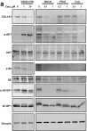
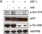
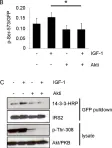
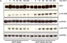
In Open Biol on 24 October 2018 by Sefried, S., Häring, H. U., et al.
Fig.3.A

-
WB
-
Collected and cropped from Open Biol by CiteAb, provided under a CC-BY license
Image 1 of 7
In Oncotarget on 15 March 2016 by Minna, E., Romeo, P., et al.
Fig.5.F

-
WB
-
Collected and cropped from Oncotarget by CiteAb, provided under a CC-BY license
Image 1 of 7
In Oncotarget on 26 January 2016 by Vergani, E., Di Guardo, L., et al.
Fig.2.C

-
WB
-
Homo sapiens (Human)
Collected and cropped from Oncotarget by CiteAb, provided under a CC-BY license
Image 1 of 7
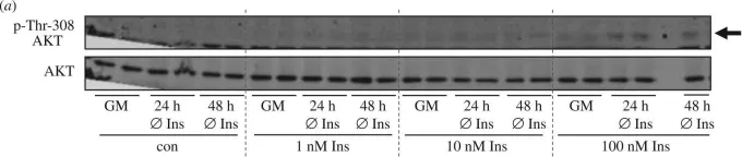
In PLoS One on 23 August 2012 by Neukamm, S. S., Toth, R., et al.
Fig.6.A

-
WB
-
Collected and cropped from PLoS One by CiteAb, provided under a CC-BY license
Image 1 of 7
In PLoS One on 23 August 2012 by Neukamm, S. S., Toth, R., et al.
Fig.6.B

-
WB
-
Collected and cropped from PLoS One by CiteAb, provided under a CC-BY license
Image 1 of 7
In PLoS One on 23 August 2012 by Neukamm, S. S., Toth, R., et al.
Fig.8.A

-
WB
-
Collected and cropped from PLoS One by CiteAb, provided under a CC-BY license
Image 1 of 7