
Copy number variations (CNVs) play a vital role in regulating genes expression and tumorigenesis. We explored the copy number alterations in early-stage lung adenocarcinoma using high-throughput sequencing and nucleic acid flight mass spectrometry technology, and found that 8q22.1-22.2 is frequently amplified in lung adenocarcinoma tissues. COX6C localizes on the region and its expression is notably enhanced that driven by amplification in lung adenocarcinoma. Knockdown of COX6C significantly inhibits the cell proliferation, and induces S-G2/M cell cycle arrest, mitosis deficiency and apoptosis. Moreover, COX6C depletion causes a deficiency in mitochondrial fusion, and impairment of oxidative phosphorylation. Mechanistically, COX6C-induced mitochondrial deficiency stimulates ROS accumulation and activates AMPK pathway, then leading to abnormality in spindle formation and chromosome segregation, activating spindle assemble checkpoint, causing mitotic arrest, and ultimately inducing cell apoptosis. Collectively, we suggested that copy amplification-mediated COX6C upregulation might serves as a prospective biomarker for prognosis and targeting therapy in patients with lung adenocarcinoma.
© 2024. The Author(s).
Product Citations: 27
In Cell Death & Disease on 19 January 2024 by Liu, S., Shao, F., et al.
-
Cancer Research
-
Cell Biology
AurkA nuclear localization is promoted by TPX2 and counteracted by protein degradation.
In Life Science Alliance on 1 May 2023 by Asteriti, I. A., Polverino, F., et al.
The AurkA kinase is a well-known mitotic regulator, frequently overexpressed in tumors. The microtubule-binding protein TPX2 controls AurkA activity, localization, and stability in mitosis. Non-mitotic roles of AurkA are emerging, and increased nuclear localization in interphase has been correlated with AurkA oncogenic potential. Still, the mechanisms leading to AurkA nuclear accumulation are poorly explored. Here, we investigated these mechanisms under physiological or overexpression conditions. We observed that AurkA nuclear localization is influenced by the cell cycle phase and nuclear export, but not by its kinase activity. Importantly, AURKA overexpression is not sufficient to determine its accumulation in interphase nuclei, which is instead obtained when AURKA and TPX2 are co-overexpressed or, to a higher extent, when proteasome activity is impaired. Expression analyses show that AURKA, TPX2, and the import regulator CSE1L are co-overexpressed in tumors. Finally, using MCF10A mammospheres we show that TPX2 co-overexpression drives protumorigenic processes downstream of nuclear AurkA. We propose that AURKA/TPX2 co-overexpression in cancer represents a key determinant of AurkA nuclear oncogenic functions.
© 2023 Asteriti et al.
-
ICC-IF
-
Homo sapiens (Human)
In Journal of Medicinal Chemistry on 23 February 2023 by Saint-Dizier, F., Matthews, T. P., et al.
The existence of multiple centrosomes in some cancer cells can lead to cell death through the formation of multipolar mitotic spindles and consequent aberrant cell division. Many cancer cells rely on HSET (KIFC1) to cluster the extra centrosomes into two groups to mimic the bipolar spindle formation of non-centrosome-amplified cells and ensure their survival. Here, we report the discovery of a novel 2-(3-benzamidopropanamido)thiazole-5-carboxylate with micromolar in vitro inhibition of HSET (KIFC1) through high-throughput screening and its progression to ATP-competitive compounds with nanomolar biochemical potency and high selectivity against the opposing mitotic kinesin Eg5. Induction of the multipolar phenotype was shown in centrosome-amplified human cancer cells treated with these inhibitors. In addition, a suitable linker position was identified to allow the synthesis of both fluorescent- and trans-cyclooctene (TCO)-tagged probes, which demonstrated direct compound binding to the HSET protein and confirmed target engagement in cells, through a click-chemistry approach.
-
Chemistry
A kinase-independent function for AURORA-A in replisome assembly during DNA replication initiation.
In Nucleic Acids Research on 20 August 2020 by Guarino Almeida, E., Renaudin, X., et al.
The catalytic activity of human AURORA-A kinase (AURKA) regulates mitotic progression, and its frequent overexpression in major forms of epithelial cancer is associated with aneuploidy and carcinogenesis. Here, we report an unexpected, kinase-independent function for AURKA in DNA replication initiation whose inhibition through a class of allosteric inhibitors opens avenues for cancer therapy. We show that genetic depletion of AURKA, or its inhibition by allosteric but not catalytic inhibitors, blocks the G1-S cell cycle transition. A catalytically inactive AURKA mutant suffices to overcome this block. We identify a multiprotein complex between AURKA and the replisome components MCM7, WDHD1 and POLD1 formed during G1, and demonstrate that allosteric but not catalytic inhibitors prevent the chromatin assembly of functional replisomes. Indeed, allosteric but not catalytic AURKA inhibitors sensitize cancer cells to inhibition of the CDC7 kinase subunit of the replication-initiating factor DDK. Thus, our findings define a mechanism essential for replisome assembly during DNA replication initiation that is vulnerable to inhibition as combination therapy in cancer.
© The Author(s) 2020. Published by Oxford University Press on behalf of Nucleic Acids Research.
-
WB
-
Biochemistry and Molecular biology
-
Genetics
Excess TPX2 Interferes with Microtubule Disassembly and Nuclei Reformation at Mitotic Exit.
In Cells on 6 February 2020 by Naso, F. D., Sterbini, V., et al.
The microtubule-associated protein TPX2 is a key mitotic regulator that contributes through distinct pathways to spindle assembly. A well-characterised function of TPX2 is the activation, stabilisation and spindle localisation of the Aurora-A kinase. High levels of TPX2 are reported in tumours and the effects of its overexpression have been investigated in cancer cell lines, while little is known in non-transformed cells. Here we studied TPX2 overexpression in hTERT RPE-1 cells, using either the full length TPX2 or a truncated form unable to bind Aurora-A, to identify effects that are dependent-or independent-on its interaction with the kinase. We observe significant defects in mitotic spindle assembly and progression through mitosis that are more severe when overexpressed TPX2 is able to interact with Aurora-A. Furthermore, we describe a peculiar, and Aurora-A-interaction-independent, phenotype in telophase cells, with aberrantly stable microtubules interfering with nuclear reconstitution and the assembly of a continuous lamin B1 network, resulting in daughter cells displaying doughnut-shaped nuclei. Our results using non-transformed cells thus reveal a previously uncharacterised consequence of abnormally high TPX2 levels on the correct microtubule cytoskeleton remodelling and G1 nuclei reformation, at the mitosis-to-interphase transition.
-
IF
-
PLA
-
ICC-IF
-
Homo sapiens (Human)
-
Cell Biology
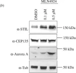
In Open Biol on 1 February 2018 by Arquint, C., Cubizolles, F., et al.
Fig.1.B

-
WB
-
Homo sapiens (Human)
Collected and cropped from Open Biol by CiteAb, provided under a CC-BY license
Image 1 of 2
In Open Biol on 1 February 2018 by Arquint, C., Cubizolles, F., et al.
Fig.1.C

-
WB
-
Homo sapiens (Human)
Collected and cropped from Open Biol by CiteAb, provided under a CC-BY license
Image 1 of 2