Vascular smooth muscle cells (VSMCs) normally exhibit a very low proliferative rate. Vessel injury triggers VSMC proliferation, in part, through focal adhesion kinase (FAK) activation, which increases transcription of cyclin D1, a key activator for cell cycle-dependent kinases (CDKs). At the same time, we also observe that FAK regulates the expression of the CDK inhibitors (CDKIs) p27 and p21. However, the mechanism of how FAK controls CDKIs in cell cycle progression is not fully understood.
We found that pharmacological and genetic FAK inhibition increased p27 and p21 by reducing stability of S-phase kinase-associated protein 2 (Skp2), which targets theCDKIs for degradation. FAK N-terminal domain interacts with Skp2 and an APC/C E3 ligase activator fizzy-related 1 (Fzr1) in the nucleus, which promote ubiquitination and degradation of both Skp2 and Fzr1. Notably, overexpression of cyclin D1 alone failed to promote proliferation of genetic FAK kinase-dead (KD) VSMCs, suggesting that the FAK-Skp2-CDKI signalling axis is distinct from the FAK-cyclin D1 pathway. However, overexpression of both cyclin D1 and Skp2 enabled proliferation of FAK-KD VSMCs, implicating that FAK ought to control both activating and inhibitory switches for CDKs. In vivo, wire injury activated FAK in the cytosol, which increased Skp2 and decreased p27 and p21 levels.
Both pharmacological FAK and genetic FAK inhibition reduced Skp2 expression in VSMCs upon injury, which significantly reduced intimal hyperplasia through elevated expression of p27 and p21. This study revealed that nuclear FAK-Skp2-CDKI signalling negatively regulates CDK activity in VSMC proliferation.
Published on behalf of the European Society of Cardiology. All rights reserved. © The Author(s) 2021. For permissions, please email: journals.permissions@oup.com.
Product Citations: 22
In Cardiovascular Research on 16 March 2022 by Jeong, K., Murphy, J. M., et al.
-
Cardiovascular biology
Requirement for Serine-384 in Caspase-2 processing and activity.
In Cell Death & Disease on 3 October 2020 by Zamaraev, A. V., Volik, P. I., et al.
Caspase-2 is a unique and conservative cysteine protease which plays an important role in several cellular processes including apoptotic cell death. Although the molecular mechanisms of its activation remain largely unclear, a major role belongs to the architecture of the caspase-2 active center. We demonstrate that the substitution of the putative phosphorylation site of caspase-2, Serine-384 to Alanine, blocks caspase-2 processing and decreases its enzymatic activity. Strikingly, in silico analysis using molecular dynamics simulations has shown that Serine-384 is crucially involved in interactions within the caspase-2 active center. It stabilizes Arginine-378, which forms a crucial hydrogen bond with the aspartate residue of a substrate. Hence, Serine-384 is essential for supporting a proper architecture of the active center of caspase-2. Moreover, molecular modeling strongly proved steric inaccessibility of Ser-384 to be phosphorylated. Importantly, a multiple alignment has demonstrated that both Serine-384 and Arg-378 residues are highly conservative across all members of caspase family, which allows us to suggest that this diade is indispensable for caspase processing and activity. Spontaneous mutations in this diade might influence oncosuppressive function of caspases, in particular of caspase-2. Likewise, the mutation of Ser-384 is associated with the development of lung squamous cell carcinoma and adenocarcinoma. Taken together, we have uncovered a central feature of the caspase-2 activation mechanism which is crucial for the regulation of its signaling network.
-
WB
-
Cell Biology
YM155 enhances the cytotoxic activity of etoposide against canine osteosarcoma cells.
In The Journal of Veterinary Medical Science / the Japanese Society of Veterinary Science on 24 August 2019 by Ong, S. M., Saeki, K., et al.
Canine osteosarcoma (OSA) is an aggressive and highly malignant primary bone tumor. Its poor survival outcome remains problematic despite recent advances in anti-cancer therapy, therefore highlighting the need for alternative treatment options or drug repositioning. The aim of this study was to determine if YM155, a small-molecule survivin inhibitor, potentiates the chemotherapeutic efficacy of etoposide against canine OSA in vitro and in vivo. In cell culture, YM155 enhanced the cytotoxic effect of etoposide against canine OSA cell lines; however, the molecular mechanism behind this effect was heterogeneous, as only one cell line had an elevated apoptotic level. In addition, this effect was not associated with survivin suppression in two of the cell lines. These results suggest that the molecular target of YM155 is not restricted to survivin alone. When tested on a murine xenograft model, the average tumor volume of the combination treatment group (YM155, 5 mg/kg, intraperitoneally, 5 consecutive days/week; and etoposide, 20 mg/kg, intraperitoneally, every 5 days) was 66% smaller than the control group, although this difference was not statistically significant (P=0.17). Further studies to improve the treatment protocol are necessary to confirm the findings of this study.
-
WB
-
Canis lupus familiaris (Domestic dog)
-
Cancer Research
-
Veterinary Research
In BMC Complementary and Alternative Medicine on 4 December 2018 by Matsushita, Y., Furutani, Y., et al.
Pancreatic cancer is one of the most aggressive human malignancies. The development of a novel drug to treat pancreatic cancer is imperative, and it is thought that complementary and alternative medicine (CAM) could yield such a candidate. Agaricus blazei Murrill is a CAM that has been tested as an anticancer drug, but its efficacy against pancreatic cancer is poorly understood. To study the potential of A. blazei in the treatment of pancreatic cancer, we examined the effects of its hot water extract on the proliferation and global gene expression profile of human pancreatic cancer cells.
Three distinct human pancreatic cancer cell lines, MIAPaCa-2, PCI-35, and PK-8, and the immortalized human pancreatic duct-epithelial cell line, HPDE, were employed. The cells were incubated with the appropriate growth medium supplemented with the hot water extract of A. blazei at final concentrations of 0.005, 0.015%, or 0.045%, and cellular proliferation was assessed for five consecutive days using an MTT assay. Apoptosis was examined by using flow cytometry and the terminal deoxynucleotidyl transferase dUTP nick-end labeling (TUNEL) assay. Caspase-dependent apoptosis was assayed using immunoblotting. Global gene expression profiles were examined using a whole human genome 44 K microarray, and the microarray results were validated by using real-time reverse transcription PCR.
The hot water extract of A. blazei significantly inhibited the proliferation of cultured pancreatic cancer cells through the induction of G0/G1 cell cycle arrest and caspase-dependent apoptosis; the effect was the smallest in HPDE cells. Furthermore, significant alterations in the global gene expression profiles of pancreatic cancer cells occurred following treatment with the hot water extract of A. blazei. Genes associated with kinetochore function, spindle formation, and centromere maintenance were particularly affected, as well as cyclins and cyclin-dependent kinases that are essential for cell cycle progression. In addition, proapoptotic genes were upregulated.
The hot water extract of A. blazei may be useful for the treatment of pancreatic cancer and is a potential candidate for the isolation of novel, active compounds specific for mitotic spindle dysfunction.
-
Cancer Research
In Cell Research on 1 August 2017 by Magadum, A., Ding, Y., et al.
Zebrafish can efficiently regenerate their heart through cardiomyocyte proliferation. In contrast, mammalian cardiomyocytes stop proliferating shortly after birth, limiting the regenerative capacity of the postnatal mammalian heart. Therefore, if the endogenous potential of postnatal cardiomyocyte proliferation could be enhanced, it could offer a promising future therapy for heart failure patients. Here, we set out to systematically identify small molecules triggering postnatal cardiomyocyte proliferation. By screening chemical compound libraries utilizing a Fucci-based system for assessing cell cycle stages, we identified carbacyclin as an inducer of postnatal cardiomyocyte proliferation. In vitro, carbacyclin induced proliferation of neonatal and adult mononuclear rat cardiomyocytes via a peroxisome proliferator-activated receptor δ (PPARδ)/PDK1/p308Akt/GSK3β/β-catenin pathway. Inhibition of PPARδ reduced cardiomyocyte proliferation during zebrafish heart regeneration. Notably, inducible cardiomyocyte-specific overexpression of constitutively active PPARδ as well as treatment with PPARδ agonist after myocardial infarction in mice induced cell cycle progression in cardiomyocytes, reduced scarring, and improved cardiac function. Collectively, we established a cardiomyocyte proliferation screening system and present a new drugable target with promise for the treatment of cardiac pathologies caused by cardiomyocyte loss.
-
WB
-
Cardiovascular biology
-
Cell Biology
In Retrovirology on 11 August 2009 by Jiang, S., Inada, T., et al.
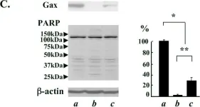
Fig.1.C

-
WB
-
Mus musculus (House mouse)
Collected and cropped from Retrovirology by CiteAb, provided under a CC-BY license
Image 1 of 1