Many devastating neuromuscular diseases currently lack effective treatments. This is in part due to a lack of drug discovery platforms capable of assessing complex human neuromuscular disease phenotypes in a scalable manner. A major obstacle has been generating scaffolds to stabilise mature contractile myofibers in a multi-well assay format amenable to high content image (HCI) analysis. This study describes the development of a scalable human induced pluripotent stem cell (iPSC)-neuromuscular disease model, whereby suspended elastomer nanofibers support long-term stability, alignment, maturation, and repeated contractions of iPSC-myofibers, innervated by iPSC-motor neurons in 96-well assay plates. In this platform, optogenetic stimulation of the motor neurons elicits robust myofiber-contractions, providing a functional readout of neuromuscular transmission. Additionally, HCI analysis provides rapid and automated quantification of axonal outgrowth, myofiber morphology, and neuromuscular synapse number and morphology. By incorporating amyotrophic lateral sclerosis (ALS)-related TDP-43G298Smutant motor neurons and CRISPR-corrected controls, key neuromuscular disease phenotypes are recapitulated, including weaker myofiber contractions, reduced axonal outgrowth, and reduced number of neuromuscular synapses. Treatment with a candidate ALS drug, the receptor-interacting protein kinase-1 (RIPK1)-inhibitor necrostatin-1, rescues these phenotypes in a dose-dependent manner, highlighting the potential of this platform to screen novel treatments for neuromuscular diseases.
Creative Commons Attribution license.
Product Citations: 10
In Biofabrication on 5 September 2023 by Cheesbrough, A., Harley, P., et al.
-
ICC-IF
-
Stem Cells and Developmental Biology
In Cells on 11 July 2020 by Lehka, L., Topolewska, M., et al.
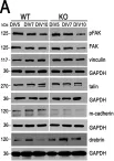
We have previously postulated that unconventional myosin VI (MVI) could be involved in myoblast differentiation. Here, we addressed the mechanism(s) of its involvement using primary myoblast culture derived from the hindlimb muscles of Snell's waltzer mice, the natural MVI knockouts (MVI-KO). We observed that MVI-KO myotubes were formed faster than control heterozygous myoblasts (MVI-WT), with a three-fold increase in the number of myosac-like myotubes with centrally positioned nuclei. There were also changes in the levels of the myogenic transcription factors Pax7, MyoD and myogenin. This was accompanied by changes in the actin cytoskeleton and adhesive structure organization. We observed significant decreases in the levels of proteins involved in focal contact formation, such as talin and focal adhesion kinase (FAK). Interestingly, the levels of proteins involved in intercellular communication, M-cadherin and drebrin, were also affected. Furthermore, time-dependent alterations in the levels of the key proteins for myoblast membrane fusion, myomaker and myomerger, without effect on their cellular localization, were observed. Our data indicate that in the absence of MVI, the mechanisms controlling cytoskeleton organization, as well as myoblast adhesion and fusion, are dysregulated, leading to the formation of aberrant myotubes.
-
WB
-
Cell Biology
PIP3 depletion rescues myoblast fusion defects in human rhabdomyosarcoma cells.
In Journal of Cell Science on 28 April 2020 by Lian, Y. L., Chen, K. W., et al.
Myoblast fusion is required for myotube formation during myogenesis, and defects in myoblast differentiation and fusion have been implicated in a number of diseases, including human rhabdomyosarcoma. Although transcriptional regulation of the myogenic program has been studied extensively, the mechanisms controlling myoblast fusion remain largely unknown. This study identified and characterized the dynamics of a distinct class of blebs, termed bubbling blebs, which are smaller than those that participate in migration. The formation of these bubbling blebs occurred during differentiation and decreased alongside a decline in phosphatidylinositol-(3,4,5)-trisphosphate (PIP3) at the plasma membrane before myoblast fusion. In a human rhabdomyosarcoma-derived (RD) cell line that exhibits strong blebbing dynamics and myoblast fusion defects, PIP3 was constitutively abundant on the membrane during myogenesis. Targeting phosphatase and tensin homolog (PTEN) to the plasma membrane reduced PIP3 levels, inhibited bubbling blebs and rescued myoblast fusion defects in RD cells. These findings highlight the differential distribution and crucial role of PIP3 during myoblast fusion and reveal a novel mechanism underlying myogenesis defects in human rhabdomyosarcoma.
© 2020. Published by The Company of Biologists Ltd.
-
IF
-
Cancer Research
-
Cell Biology
In Muscle Nerve on 1 May 2019 by Verma, M., Asakura, Y., et al.
The vasculature and blood flow in muscle are perturbed in Duchenne muscular dystrophy (DMD) and its mdx mouse model. MicroRNA-92a (miR-92a) is enriched in endothelial cells, especially during ischemic injury.
Because antagonizing miR-92a was shown to result in increased proliferation and migration of endothelial cells and recovery from ischemia, we assessed the effects of Antagomir-92a in vitro in muscle stem cell culture and in vivo in mdx mice.
miR-92a was found to be highly expressed in muscle endothelial cells and satellite cells. Treatment with Antagomir-92a increased capillary density and tissue perfusion, which was accompanied by an increase in satellite cells. However, Antagomir-92a-treated mdx mice showed no histological improvement and had worse muscle function. Antagomir-92a suppressed myogenic differentiation in satellite cell culture.
AntagomiR-92a improves the vasculature but not the muscle in mdx mice, possibly due to its side effects on satellite cell differentiation. Muscle Nerve 59:594-594, 2019.
© 2019 Wiley Periodicals, Inc.
-
Cardiovascular biology
Maintaining bovine satellite cells stemness through p38 pathway.
In Scientific Reports on 17 July 2018 by Ding, S., Swennen, G. N. M., et al.
Isolating and maintaining the appropriate stem cell for large scale cell culture is essential in tissue engineering or food production. For bovine satellite cells an optimized isolation and purification protocol is lacking and there is also no detailed understanding on the factors that maintain stemness of these cells. Here, we set up a fluorescence-activated cell sorting strategy to enrich bovine satellite cells. We found that p38-MAPK signalling is activated and PAX7 expression is gradually lost during satellite cell proliferation. The p38 inhibitor (SB203580) treatment maintained PAX7 expression but inhibited the fusion of satellite cells in a concentration-dependent way in short-term incubation. The mechanism of p38 inhibition was confirmed by inhibiting canonical p38 signalling, i.e. HSP27. Long-term culture with an appropriate concentration of p38i enhanced the proliferation and PAX7 expression, while the differentiation capacity recovered and was enhanced compared to vehicle control. These studies indicate that bovine satellite cells maintenance depends on cell purity and p38 MAPK signalling. Inhibition of p38 MAPK signaling is a promising strategy to facilitate large scale cell expansion of primary cells for tissue engineering and cultured meat purposes.
-
IF
-
ICC-IF
-
Veterinary Research
In Cells on 11 July 2020 by Lehka, L., Topolewska, M., et al.
Fig.3.A

-
WB
-
Collected and cropped from Cells by CiteAb, provided under a CC-BY license
Image 1 of 1