The Golgi apparatus is the major sorting hub in the secretory pathway and particularly important for protein sorting in neurons. Knowledge about protein localization in Golgi compartments is largely based on work in cell lines. Here, we systematically compared protein localization of 21 endogenous proteins in the Golgi apparatus of mouse neurons using confocal microscopy and line scan analysis. We localized these proteins by measuring the distance relative to the canonical TGN marker TGN38. Based on this, proteins fell into three groups: upstream of, overlapping with or downstream of TGN38. Seven proteins showed complete overlap with TGN38, while proteins downstream of TGN38 were located at varying distances from TGN38. Proteins upstream of TGN38 were localized in between TGN38 and the cis-/medial Golgi markers Giantin and GM130. This localization was consistent with protein function. Our data provide an overview of the relative localization of endogenous proteins in the Golgi of primary mouse neurons.
© 2023. The Author(s).
Product Citations: 29
Mapping localization of 21 endogenous proteins in the Golgi apparatus of rodent neurons.
In Scientific Reports on 18 February 2023 by van Bommel, D. M., Toonen, R. F., et al.
-
ICC-IF
-
Mus musculus (House mouse)
-
Cell Biology
-
Neuroscience
Synaptotagmin-11 inhibits spontaneous neurotransmission through vti1a.
In Journal of Neurochemistry on 1 November 2021 by Li, W. R., Wang, Y. L., et al.
Recent work has revealed that spontaneous release plays critical roles in the central nervous system, but how it is regulated remains elusive. Here, we report that synaptotagmin-11 (Syt11), a Ca2+ -independent Syt isoform associated with schizophrenia and Parkinson's disease, suppressed spontaneous release. Syt11-knockout hippocampal neurons showed an increased frequency of miniature excitatory post-synaptic currents while over-expression of Syt11 inversely decreased the frequency. Neither knockout nor over-expression of Syt11 affected the average amplitude, suggesting the pre-synaptic regulation of spontaneous neurotransmission by Syt11. Glutathione S-transferase pull-down, co-immunoprecipitation, and affinity-purification experiments demonstrated a direct interaction of Syt11 with vps10p-tail-interactor-1a (vti1a), a non-canonical SNARE protein that maintains spontaneous release. Importantly, knockdown of vti1a reversed the phenotype of Syt11 knockout, identifying vti1a as the main target of Syt11 inhibition. Domain analysis revealed that the C2A domain of Syt11 bound vti1a with high affinity. Consistently, expression of the C2A domain alone rescued the phenotype of elevated spontaneous release in Syt11-knockout neurons similar to the full-length protein. Altogether, our results suggest that Syt11 inhibits vti1a-containing vesicles during spontaneous release.
© 2021 International Society for Neurochemistry.
-
Neuroscience
Control of synaptic vesicle release probability via VAMP4 targeting to endolysosomes.
In Science Advances on 1 April 2021 by Ivanova, D., Dobson, K. L., et al.
Synaptic vesicle (SV) release probability (Pr), determines the steady state and plastic control of neurotransmitter release. However, how diversity in SV composition arises and regulates the Pr of individual SVs is not understood. We found that modulation of the copy number of the noncanonical vesicular SNARE (soluble N-ethylmaleimide-sensitive factor attachment protein receptor), vesicle-associated membrane protein 4 (VAMP4), on SVs is key for regulating Pr. Mechanistically, this is underpinned by its reduced ability to form an efficient SNARE complex with canonical plasma membrane SNAREs. VAMP4 has unusually high synaptic turnover and is selectively sorted to endolysosomes during activity-dependent bulk endocytosis. Disruption of endolysosomal trafficking and function markedly increased the abundance of VAMP4 in the SV pool and inhibited SV fusion. Together, our results unravel a new mechanism for generating SV heterogeneity and control of Pr through coupling of SV recycling to a major clearing system that regulates protein homeostasis.
Copyright © 2021 The Authors, some rights reserved; exclusive licensee American Association for the Advancement of Science. No claim to original U.S. Government Works. Distributed under a Creative Commons Attribution License 4.0 (CC BY).
-
WB
-
Mus musculus (House mouse)
OPTN recruitment to a Golgi-proximal compartment regulates immune signalling and cytokine secretion.
In Journal of Cell Science on 15 June 2020 by O'Loughlin, T., Kruppa, A. J., et al.
Optineurin (OPTN) is a multifunctional protein involved in autophagy and secretion, as well as nuclear factor κB (NF-κB) and IRF3 signalling, and OPTN mutations are associated with several human diseases. Here, we show that, in response to viral RNA, OPTN translocates to foci in the perinuclear region, where it negatively regulates NF-κB and IRF3 signalling pathways and downstream pro-inflammatory cytokine secretion. These OPTN foci consist of a tight cluster of small membrane vesicles, which are positive for ATG9A. Disease mutations in OPTN linked to primary open-angle glaucoma (POAG) cause aberrant foci formation in the absence of stimuli, which correlates with the ability of OPTN to inhibit signalling. By using proximity labelling proteomics, we identify the linear ubiquitin assembly complex (LUBAC), CYLD and TBK1 as part of the OPTN interactome and show that these proteins are recruited to this OPTN-positive perinuclear compartment. Our work uncovers a crucial role for OPTN in dampening NF-κB and IRF3 signalling through the sequestration of LUBAC and other positive regulators in this viral RNA-induced compartment, leading to altered pro-inflammatory cytokine secretion.
© 2020. Published by The Company of Biologists Ltd.
-
Cell Biology
-
Immunology and Microbiology
DYRK3-Controlled Phase Separation Organizes the Early Secretory Pathway
Preprint on BioRxiv : the Preprint Server for Biology on 10 February 2020 by Gallo, R., Rai, A., et al.
h4>Summary/h4> The dual-specificity kinase DYRK3 controls formation and dissolution of several intracellular condensates thereby regulating various cell physiological processes. Here we report that DYRK3 establishes a dynamic equilibrium between condensation and dissolution of proteins associated with membranous structures of the early secretory pathway to organize membrane traffic between the ER and the Golgi complex in mammalian cells. This depends on the peripheral membrane protein Sec16A, whose N-terminal disordered region forms DYRK3-controlled liquid-like condensates on the surface of the ER and co-phase separates with multiple ER exit site components and a subset of matrix proteins specifically associated with ERGIC and cis-Golgi. Our findings support a mechanism whereby multiple interacting and differentially regulated intracellular condensates create favorable environments for directional membrane traffic in eukaryotic cells.
In Sci Adv on 1 April 2021 by Ivanova, D., Dobson, K. L., et al.
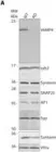
Fig.6.A

-
WB
-
Mus musculus (House mouse)
Collected and cropped from Sci Adv by CiteAb, provided under a CC-BY license
Image 1 of 2
In Nat Commun on 24 August 2018 by Emperador-Melero, J., Huson, V., et al.
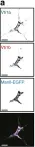
Fig.6.A

-
ICC-IF
-
Mus musculus (House mouse)
Collected and cropped from Nat Commun by CiteAb, provided under a CC-BY license
Image 1 of 2