Autophagy initiation is regulated by the ULK1 kinase complex. To gain insights into functions of the holo-complex, we generated a deep interactome by combining affinity purification- and proximity labeling-mass spectrometry of all four complex members: ULK1, ATG13, ATG101, and RB1CC1/FIP200. Under starvation conditions, the ULK1 complex interacts with several protein and lipid kinases and phosphatases, implying the formation of a signalosome. Interestingly, several selective autophagy receptors also interact with ULK1, indicating the activation of selective autophagy pathways by nutrient starvation. One effector of the ULK1 complex is the HSC/HSP70 co-chaperone BAG2, which regulates the subcellular localization of the VPS34 lipid kinase complex member AMBRA1. Depending on the nutritional status, BAG2 has opposing roles. In growth conditions, the unphosphorylated form of BAG2 sequesters AMBRA1, attenuating autophagy induction. In starvation conditions, ULK1 phosphorylates BAG2 on Ser31, which supports the recruitment of AMBRA1 to the ER membrane, positively affecting autophagy.
Copyright © 2024 The Author(s). Published by Elsevier Inc. All rights reserved.
Product Citations: 48
The ULK1 effector BAG2 regulates autophagy initiation by modulating AMBRA1 localization.
In Cell Reports on 24 September 2024 by Sankar, D. S., Kaeser-Pebernard, S., et al.
-
Cell Biology
Atlastin-1 regulates endosomal tubulation and lysosomal proteolysis in human cortical neurons.
In Neurobiology of Disease on 1 September 2024 by Zlámalová, E., Rodger, C., et al.
Mutation of the ATL1 gene is one of the most common causes of hereditary spastic paraplegia (HSP), a group of genetic neurodegenerative conditions characterised by distal axonal degeneration of the corticospinal tract axons. Atlastin-1, the protein encoded by ATL1, is one of three mammalian atlastins, which are homologous dynamin-like GTPases that control endoplasmic reticulum (ER) morphology by fusing tubules to form the three-way junctions that characterise ER networks. However, it is not clear whether atlastin-1 is required for correct ER morphology in human neurons and if so what the functional consequences of lack of atlastin-1 are. Using CRISPR-inhibition we generated human cortical neurons lacking atlastin-1. We demonstrate that ER morphology was altered in these neurons, with a reduced number of three-way junctions. Neurons lacking atlastin-1 had longer endosomal tubules, suggestive of defective tubule fission. This was accompanied by reduced lysosomal proteolytic capacity. As well as demonstrating that atlastin-1 is required for correct ER morphology in human neurons, our results indicate that lack of a classical ER-shaping protein such as atlastin-1 may cause altered endosomal tubulation and lysosomal proteolytic dysfunction. Furthermore, they strengthen the idea that defective lysosome function contributes to the pathogenesis of a broad group of HSPs, including those where the primary localisation of the protein involved is not at the endolysosomal system.
Copyright © 2024 The Authors. Published by Elsevier Inc. All rights reserved.
-
Cell Biology
-
Neuroscience
Mechanism and regulation of cargo entry into the Commander endosomal recycling pathway.
In Nature Communications on 21 August 2024 by Butkovič, R., Walker, A. P., et al.
Commander is a multiprotein complex that orchestrates endosomal recycling of integral cargo proteins and is essential for normal development. While the structure of this complex has recently been described, how cargo proteins are selected for Commander-mediated recycling remains unclear. Here we identify the mechanism through which the unstructured carboxy-terminal tail of the cargo adaptor sorting nexin-17 (SNX17) directly binds to the Retriever sub-complex of Commander. SNX17 adopts an autoinhibited conformation where its carboxy-terminal tail occupies the cargo binding groove. Competitive cargo binding overcomes this autoinhibition, promoting SNX17 endosomal residency and the release of the tail for Retriever association. Furthermore, our study establishes the central importance of SNX17-Retriever association in the handover of integrin and lipoprotein receptor cargoes into pre-existing endosomal retrieval sub-domains. In describing the principal mechanism of cargo entry into the Commander recycling pathway we provide key insight into the function and regulation of this evolutionary conserved sorting pathway.
© 2024. The Author(s).
-
Cell Biology
In Molecular Biology of the Cell on 1 March 2024 by van der Beek, J., de Heus, C., et al.
The multisubunit HOPS tethering complex is a well-established regulator of lysosome fusion with late endosomes and autophagosomes. However, the role of the HOPS complex in other stages of endo-lysosomal trafficking is not well understood. To address this, we made HeLa cells knocked out for the HOPS-specific subunits Vps39 or Vps41, or the HOPS-CORVET-core subunits Vps18 or Vps11. In all four knockout cells, we found that endocytosed cargos were trapped in enlarged endosomes that clustered in the perinuclear area. By correlative light-electron microscopy, these endosomes showed a complex ultrastructure and hybrid molecular composition, displaying markers for early (Rab5, PtdIns3P, EEA1) as well as late (Rab7, CD63, LAMP1) endosomes. These "HOPS bodies" were not acidified, contained enzymatically inactive cathepsins and accumulated endocytosed cargo and cation-independent mannose-6-phosphate receptor (CI-MPR). Consequently, CI-MPR was depleted from the TGN, and secretion of lysosomal enzymes to the extracellular space was enhanced. Strikingly, HOPS bodies also contained the autophagy proteins p62 and LC3, defining them as amphisomes. Together, these findings show that depletion of the lysosomal HOPS complex has a profound impact on the functional organization of the entire endosomal system and suggest the existence of a HOPS-independent mechanism for amphisome formation.
-
Cell Biology
Phosphatidylserine synthesis controls oncogenic B cell receptor signaling in B cell lymphoma.
In The Journal of Cell Biology on 5 February 2024 by Omi, J., Kato, T., et al.
Cancer cells harness lipid metabolism to promote their own survival. We screened 47 cancer cell lines for survival dependency on phosphatidylserine (PS) synthesis using a PS synthase 1 (PTDSS1) inhibitor and found that B cell lymphoma is highly dependent on PS. Inhibition of PTDSS1 in B cell lymphoma cells caused a reduction of PS and phosphatidylethanolamine levels and an increase of phosphoinositide levels. The resulting imbalance of the membrane phospholipidome lowered the activation threshold for B cell receptor (BCR), a B cell-specific survival mechanism. BCR hyperactivation led to aberrant elevation of downstream Ca2+ signaling and subsequent apoptotic cell death. In a mouse xenograft model, PTDSS1 inhibition efficiently suppressed tumor growth and prolonged survival. Our findings suggest that PS synthesis may be a critical vulnerability of malignant B cell lymphomas that can be targeted pharmacologically.
© 2023 Omi et al.
-
Cancer Research
-
Cell Biology
-
Immunology and Microbiology
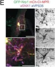
In Proc Natl Acad Sci U S A on 21 June 2022 by Simonetti, B., Daly, J. L., et al.
Fig.3.E

-
IHC-IF
-
Collected and cropped from Proc Natl Acad Sci U S A by CiteAb, provided under a CC-BY license
Image 1 of 3
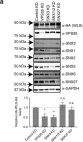
In Nat Commun on 13 September 2018 by McGough, I. J., de Groot, R. E. A., et al.
Fig.3.A

-
WB
-
Collected and cropped from Nat Commun by CiteAb, provided under a CC-BY license
Image 1 of 3
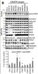
In EMBO J on 17 January 2018 by Jimenez-Orgaz, A., Kvainickas, A., et al.
Fig.3.B

-
WB
-
Homo sapiens (Human)
Collected and cropped from EMBO J by CiteAb, provided under a CC-BY license
Image 1 of 3