Protein synthesis is a process finely regulated in all cell types but specially in neurons as they need rapid changes in protein concentration for synaptic plasticity. Alterations in translation rates have been shown in diseases affecting the brain. In Huntington's disease (HD), an autosomal dominant neurodegenerative disorder characterized by the presence of motor, cognitive and psychiatric symptoms, we have shown that translation is increased in the striatum contributing to motor symptoms. However, very little is known about how translation modulates motor function in physiological conditions. To study this, we overexpressed a constitutively active mutant form of 4E-BP1 (4E-BP1F113A), a translation repressor, in the striatum of wild-type mice and performed motor tests. One month after striatal injection of adeno-associated viral vectors expressing 4E-BP1F113A, mice exhibited motor symptoms similar to those observed in the R6/1 HD mouse model. Unexpectedly, de novo protein synthesis and 4E-BP1 phosphorylation were enhanced in the striatum of wild-type mice overexpressing 4E-BP1F113A. Moreover, the striatum of these animals showed alterations in protein levels of neuronal markers similar to that observed in HD striatum. Altogether, our results indicate that enhanced protein synthesis in the striatum induces neuronal dysfunction and motor symptoms, and reinforce the idea that increased translation is involved in HD pathogenesis.
© The Author(s) 2025. Published by Oxford University Press on behalf of the Guarantors of Brain.
Product Citations: 51
Increased translation in adult mouse striatum is sufficient to induce motor dysfunction.
In Brain Communications on 7 July 2025 by Castany-Pladevall, C., Creus-Muncunill, J., et al.
-
Biochemistry and Molecular biology
In Neurobiology of Disease on 1 May 2025 by Lebouc, M., Bonamy, L., et al.
Huntington's disease (HD) is a complex neurodegenerative disorder with cognitive and motor symptoms that typically manifest in adulthood. However, embryonic brain development impairments leading to cortical defects in HD mutation carriers has been shown recently supporting a neurodevelopmental component in HD. Despite HD is primarily recognized as a striatal pathology, developmental alterations in this structure, particularly during the early postnatal period, remain poorly understood. To fill this gap, we examined striatal development in newborn R6/1 mice. We found that D2 receptor-expressing indirect-pathway medium spiny neurons (D2-MSNs) present in the matrix striatal compartment undergo early morphological and electrophysiological maturation. Altered electrophysiological properties were also observed in newborn CAG140 mice. Additionally, we also observed a D2-MSN-selective reduction in glutamatergic cortico-striatal transmission at the beginning of the second postnatal week as well as a reduced projection of D2-MSNs onto the GPe at birth in R6/1 mice. All these alterations were transient with the circuit normalizing after the second postnatal week. These results identify a compartment- and cell-type specific defect in D2-MSNs maturation, which can contribute in their latter vulnerability, as this cell-type is the first to degenerate in HD during adulthood.
Copyright © 2025 The Authors. Published by Elsevier Inc. All rights reserved.
-
Neuroscience
-
Stem Cells and Developmental Biology
In Frontiers in Molecular Neuroscience on 5 February 2025 by Fritz, M., Rosa, P. B., et al.
The neurotransmitter acetylcholine has since long been implicated in reward learning and drug addiction. However, the role of specific cholinergic receptor subtypes on different neuronal populations remain elusive. Here, we studied the function of nicotinic acetylcholinergic alpha 7 receptors (α7 nAChRs) in cocaine and food-enforced behaviors. We found that global deletion of α7 nAChRs in mice attenuates cocaine seeking in a Pavlovian conditioned place preference paradigm and decreases operant responding to cocaine in a runway task and in self-administration, without influencing responding to palatable food. This effect can be attributed to alpha 7 receptor signaling in the striatum, as selective deletion of striatal α7 nAChRs using a viral vector approach resulted in a similar decrease in cocaine-preference as that of global deletion. To investigate which type of striatal neurons are responsible for this effect, we selectively targeted Cholinergic (ChAT-expressing) neurons and dopamine D1-receptor (D1R) expressing neurons. Mice with conditional deletion of α7 nAChRs in ChAT-neurons (α7 nAChR-ChATCre) exhibited decreased cocaine place preference and intact place preference for food, while α7 nAChR-D1RCre mice had no changes in reward learning to neither food nor cocaine. Cocaine induction of striatal immediate early gene expression of cFos, FosB, Arc and EGR2 was blocked in α7 nAChR-ChATCre mice, demonstrating the importance of α7 nAChRs on cholinergic neurons for striatal neuronal activity changes. Collectively, our findings show that α7 nAChRs on cholinergic interneurons in the striatum are pivotal for learning processes related to cocaine, but not food reward.
Copyright © 2025 Fritz, Rosa, Wilhelms, Jaarola, Ruud, Engblom and Klawonn.
-
IHC-Fr-Float
-
Mus musculus (House mouse)
-
Neuroscience
Correction of RBFOX1 deficit rescues Huntington’s disease mis-splicing and pathology
Preprint on BioRxiv : the Preprint Server for Biology on 7 November 2024 by Lozano-Muñoz, D., Elorza, A., et al.
RNA mis-splicing correction therapies have been developed for neurological disorders like spinal muscular atrophy and neuronal ceroid lipofuscinosis. In Huntington’s disease (HD), pathogenic mis-splicing was initially observed in genes linked to neurodegeneration, such as HTT itself, MAPT , and TAF1 . Later, genome-wide analyses identified a broader mis-splicing signature in HD brains, involving additional neurodegeneration-related genes. Correcting each mis-spliced gene individually would be unfeasible, highlighting the need to target upstream splicing factors altered in HD. Our previous motif-enrichment analyses of intronic sequences flanking the exons mis-spliced in HD identified RBFOX and U2AF2 as candidate splicing factors, both of which are reduced in HD brains. In this study, we tested their pathogenic relevance generating conditional transgenic mouse models that overexpress RBFOX1 or U2AF2 in forebrain neurons and combining them with HD mice. Our results show that moderate overexpression of RBFOX1, but not U2AF2, corrects multiple HD-associated mis-splicing events and alleviates HD mice neuropathology and motor symptoms. These findings demonstrate that RBFOX1 downregulation contributes to HD pathology and underscore the therapeutic potential of strategies aimed at increasing RBFOX1 levels.
-
Pathology
Preprint on BioRxiv : the Preprint Server for Biology on 10 May 2024 by Lebouc, M., Bonamy, L., et al.
Huntington’s disease (HD) is an inherited neurodegenerative disorder caused by a mutation in the gene encoding the Huntingtin protein (Htt). While symptoms, primarily characterized by progressive deterioration of the striatum and motor and cognitive functions, typically manifest in adulthood, recent studies have also highlighted developmental defects in HD. Indeed, alterations in cortical and striatal development have been observed in individuals carrying the mutation as early as in embryonic stages. However, despite the striatum being one of the most affected regions in HD, few studies have investigated potential developmental alterations in this structure, especially in the early weeks after birth. To address this question, we compared striatal development between wild-type (WT) mice and two murine models of HD, R6/1 and CAG140 mice crossed with reporter mice to identify D1- and D2-expressing medium spiny neurons (D1- and D2-MSNs). Using ex vivo electrophysiology and neuronal reconstruction, we observed that the maturation of electrical properties was selectively disrupted in D2-MSNs of the matrix compartment of HD mice during the first post-natal days. D2-MSNs arbor also an increased dendritic complexity. When studying the establishment of striatal afferents, we observed that cortico-striatal glutamatergic transmission was specifically reduced in D2-MSNs during the second postnatal week. All these alterations were transient before the circuit normalized on its own after the second postnatal week. These anatomical and electrophysiological data highlight the significant impact of the Htt mutation on numerous striatal development processes during the postnatal period. Interestingly, we observed that these alterations specifically affect MSNs in the indirect pathway. This preferential vulnerability aligns with the early death of these neurons in adulthood, suggesting that early treatment of these alterations could potentially modify the disease’s progression.
-
Mus musculus (House mouse)
-
Neuroscience
-
Stem Cells and Developmental Biology
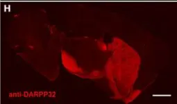
In Front Behav Neurosci on 29 April 2014 by Ting, J. T. & Feng, G.
Fig.5.H

-
IHC-IF
-
Mus musculus (House mouse)
Collected and cropped from Front Behav Neurosci by CiteAb, provided under a CC-BY license
Image 1 of 1