Nuclear entry of HIV-1 replication complexes through intact nuclear pore complexes is critical for successful infection. The host protein cleavage-and-polyadenylation-specificity-factor-6 (CPSF6) has been implicated in different stages of early HIV-1 replication. Applying quantitative microscopy of HIV-1 reverse-transcription and pre-integration-complexes (RTC/PIC), we show that CPSF6 is strongly recruited to nuclear replication complexes but absent from cytoplasmic RTC/PIC in primary human macrophages. Depletion of CPSF6 or lack of CPSF6 binding led to accumulation of HIV-1 subviral complexes at the nuclear envelope of macrophages and reduced infectivity. Two-color stimulated-emission-depletion microscopy indicated that under these circumstances HIV-1 complexes are retained inside the nuclear pore and undergo CA-multimer dependent CPSF6 clustering adjacent to the nuclear basket. We propose that nuclear entry of HIV-1 subviral complexes in macrophages is mediated by consecutive binding of Nup153 and CPSF6 to the hexameric CA lattice.
© 2019, Bejarano et al.
Product Citations: 17
In eLife on 23 January 2019 by Bejarano, D. A., Peng, K., et al.
-
IF
LEDGF/p75 Deficiency Increases Deletions at the HIV-1 cDNA Ends.
In Viruses on 15 September 2017 by Bueno, M. T. D., Reyes, D., et al.
Processing of unintegrated linear HIV-1 cDNA by the host DNA repair system results in its degradation and/or circularization. As a consequence, deficient viral cDNA integration generally leads to an increase in the levels of HIV-1 cDNA circles containing one or two long terminal repeats (LTRs). Intriguingly, impaired HIV-1 integration in LEDGF/p75-deficient cells does not result in a correspondent increase in viral cDNA circles. We postulate that increased degradation of unintegrated linear viral cDNA in cells lacking the lens epithelium-derived growth factor (LEDGF/p75) account for this inconsistency. To evaluate this hypothesis, we characterized the nucleotide sequence spanning 2-LTR junctions isolated from LEDGF/p75-deficient and control cells. LEDGF/p75 deficiency resulted in a significant increase in the frequency of 2-LTRs harboring large deletions. Of note, these deletions were dependent on the 3' processing activity of integrase and were not originated by aberrant reverse transcription. Our findings suggest a novel role of LEDGF/p75 in protecting the unintegrated 3' processed linear HIV-1 cDNA from exonucleolytic degradation.
-
WB
In Journal of Molecular Biology on 17 July 2016 by Lopez, A. P., Kugelman, J. R., et al.
The lens epithelium-derived growth factor p75 (LEDGF/p75) is a chromatin-bound protein essential for efficient lentiviral integration. Genome-wide studies have located LEDGF/p75 inside actively transcribed genes where it mediates lentiviral integration. Although its role in HIV-1 integration is clearly established, the role of LEDGF/p75-associated proteins in HIV-1 infection remains unexplored. Using protein-protein interaction assays, we demonstrated that LEDGF/p75 complexes with a chromatin-remodeling complex facilitates chromatin transcription (FACT), a heterodimer of the structure-specific recognition protein 1 (SSRP1) and the human homolog of suppressor of Ty 16 (hSpt16). Detailed analysis of the interaction of LEDGF/p75 with the FACT complex indicates that LEDGF/p75 interacts with SSRP1 in an hSpt16-independent manner that requires the PWWP domain of LEDGF proteins and the HMG domain of SSRP1. Functional characterizations demonstrate a LEDGF/p75-independent role of SSRP1 in the regulation of HIV-1 replication. shRNA-mediated partial knockdown of SSRP1 reduces HIV-1 infection, but not Murine Leukemia Virus, in human CD4(+) T cells. Similarly, SSRP1 knockdown affects infection by HIV-1-derived viruses that express genes from the viral LTR but not from an internal immediate-early CMV promoter, suggesting a role of SSRP1 in LTR-driven gene expression but not in viral DNA integration. Together, our data demonstrate for the first time the association of LEDGF proteins with the FACT complex and give further support to a role of SSRP1 in HIV-1 infection.Copyright © 2016 Elsevier Ltd. All rights reserved.
-
WB
-
Mus musculus (House mouse)
-
Biochemistry and Molecular biology
In Oncogene on 12 November 2015 by Kanu, N., Grönroos, E., et al.
Defining mechanisms that generate intratumour heterogeneity and branched evolution may inspire novel therapeutic approaches to limit tumour diversity and adaptation. SETD2 (Su(var), Enhancer of zeste, Trithorax-domain containing 2) trimethylates histone-3 lysine-36 (H3K36me3) at sites of active transcription and is mutated in diverse tumour types, including clear cell renal carcinomas (ccRCCs). Distinct SETD2 mutations have been identified in spatially separated regions in ccRCC, indicative of intratumour heterogeneity. In this study, we have addressed the consequences of SETD2 loss-of-function through an integrated bioinformatics and functional genomics approach. We find that bi-allelic SETD2 aberrations are not associated with microsatellite instability in ccRCC. SETD2 depletion in ccRCC cells revealed aberrant and reduced nucleosome compaction and chromatin association of the key replication proteins minichromosome maintenance complex component (MCM7) and DNA polymerase δ hindering replication fork progression, and failure to load lens epithelium-derived growth factor and the Rad51 homologous recombination repair factor at DNA breaks. Consistent with these data, we observe chromosomal breakpoint locations are biased away from H3K36me3 sites in SETD2 wild-type ccRCCs relative to tumours with bi-allelic SETD2 aberrations and that H3K36me3-negative ccRCCs display elevated DNA damage in vivo. These data suggest a role for SETD2 in maintaining genome integrity through nucleosome stabilization, suppression of replication stress and the coordination of DNA repair.
-
WB
-
Homo sapiens (Human)
-
Cancer Research
-
Genetics
Nuclear architecture dictates HIV-1 integration site selection.
In Nature on 14 May 2015 by Marini, B., Kertesz-Farkas, A., et al.
Long-standing evidence indicates that human immunodeficiency virus type 1 (HIV-1) preferentially integrates into a subset of transcriptionally active genes of the host cell genome. However, the reason why the virus selects only certain genes among all transcriptionally active regions in a target cell remains largely unknown. Here we show that HIV-1 integration occurs in the outer shell of the nucleus in close correspondence with the nuclear pore. This region contains a series of cellular genes, which are preferentially targeted by the virus, and characterized by the presence of active transcription chromatin marks before viral infection. In contrast, the virus strongly disfavours the heterochromatic regions in the nuclear lamin-associated domains and other transcriptionally active regions located centrally in the nucleus. Functional viral integrase and the presence of the cellular Nup153 and LEDGF/p75 integration cofactors are indispensable for the peripheral integration of the virus. Once integrated at the nuclear pore, the HIV-1 DNA makes contact with various nucleoporins; this association takes part in the transcriptional regulation of the viral genome. These results indicate that nuclear topography is an essential determinant of the HIV-1 life cycle.
-
WB
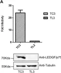
In Viruses on 15 September 2017 by Bueno, M. T. D., Reyes, D., et al.
Fig.1.A

-
WB
-
Collected and cropped from Viruses by CiteAb, provided under a CC-BY license
Image 1 of 6
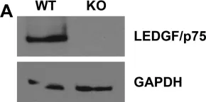
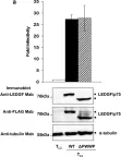
In PLoS Pathog on 1 May 2014 by Sharma, A., Slaughter, A., et al.
Fig.8.A

-
WB
-
Collected and cropped from PLoS Pathog by CiteAb, provided under a CC-BY license
Image 1 of 6
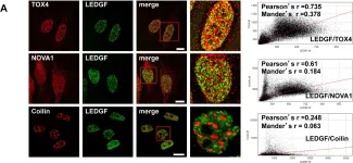
In PLoS One on 7 December 2013 by Morchikh, M., Naughtin, M., et al.
Fig.3.A

-
ICC-IF
-
Homo sapiens (Human)
Collected and cropped from PLoS One by CiteAb, provided under a CC-BY license
Image 1 of 6
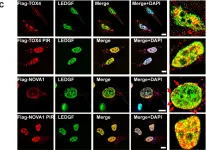
In PLoS One on 7 December 2013 by Morchikh, M., Naughtin, M., et al.
Fig.3.C

-
ICC-IF
-
Homo sapiens (Human)
Collected and cropped from PLoS One by CiteAb, provided under a CC-BY license
Image 1 of 6
In Retrovirology on 21 April 2011 by Astiazaran, P., Bueno, M. T., et al.
Fig.1.A

-
WB
-
Collected and cropped from Retrovirology by CiteAb, provided under a CC-BY license
Image 1 of 6
In Retrovirology on 21 April 2011 by Astiazaran, P., Bueno, M. T., et al.
Fig.2.A

-
WB
-
Collected and cropped from Retrovirology by CiteAb, provided under a CC-BY license
Image 1 of 6