Blood vessel formation relies on biochemical and mechanical signals, particularly during sprouting angiogenesis when endothelial tip cells (TCs) guide sprouting through filopodia formation. The contribution of BMP receptors in defining tip-cell characteristics is poorly understood. Our study combines genetic, biochemical, and molecular methods together with 3D traction force microscopy, which reveals an essential role of BMPR2 for actin-driven filopodia formation and mechanical properties of endothelial cells (ECs). Targeting of Bmpr2 reduced sprouting angiogenesis in zebrafish and BMPR2-deficient human ECs formed fewer filopodia, affecting cell migration and actomyosin localization. Spheroid assays revealed a reduced sprouting of BMPR2-deficient ECs in fibrin gels. Even more strikingly, in mosaic spheroids, BMPR2-deficient ECs failed to acquire tip-cell positions. Yet, 3D traction force microscopy revealed that these distinct cell behaviors of BMPR2-deficient tip cells cannot be explained by differences in force-induced matrix deformations, even though these cells adopted distinct cone-shaped morphologies. Notably, BMPR2 positively regulates local CDC42 activity at the plasma membrane to promote filopodia formation. Our findings reveal that BMPR2 functions as a nexus integrating biochemical and biomechanical processes crucial for TCs during angiogenesis.
© 2025. The Author(s).
Product Citations: 30
In Communications Biology on 8 January 2025 by Hiepen, C., Benamar, M., et al.
-
Homo sapiens (Human)
Control of neuronal excitation-inhibition balance by BMP-SMAD1 signalling.
In Nature on 1 May 2024 by Okur, Z., Schlauri, N., et al.
Throughout life, neuronal networks in the mammalian neocortex maintain a balance of excitation and inhibition, which is essential for neuronal computation1,2. Deviations from a balanced state have been linked to neurodevelopmental disorders, and severe disruptions result in epilepsy3-5. To maintain balance, neuronal microcircuits composed of excitatory and inhibitory neurons sense alterations in neural activity and adjust neuronal connectivity and function. Here we identify a signalling pathway in the adult mouse neocortex that is activated in response to increased neuronal network activity. Overactivation of excitatory neurons is signalled to the network through an increase in the levels of BMP2, a growth factor that is well known for its role as a morphogen in embryonic development. BMP2 acts on parvalbumin-expressing (PV) interneurons through the transcription factor SMAD1, which controls an array of glutamatergic synapse proteins and components of perineuronal nets. PV-interneuron-specific disruption of BMP2-SMAD1 signalling is accompanied by a loss of glutamatergic innervation in PV cells, underdeveloped perineuronal nets and decreased excitability. Ultimately, this impairment of the functional recruitment of PV interneurons disrupts the cortical excitation-inhibition balance, with mice exhibiting spontaneous epileptic seizures. Our findings suggest that developmental morphogen signalling is repurposed to stabilize cortical networks in the adult mammalian brain.
© 2024. The Author(s).
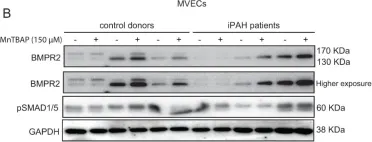
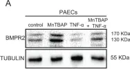
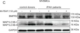
In International Journal of Molecular Sciences on 26 January 2023 by Song, Y. S., Zaitoun, I. S., et al.
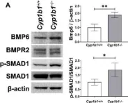
Cytochrome P450 (CYP) 1B1 is a heme-containing monooxygenase found mainly in extrahepatic tissues, including the retina. CYP1B1 substrates include exogenous aromatic hydrocarbons, such as dioxins, and endogenous bioactive compounds, including 17β-estradiol (E2) and arachidonic acid. The endogenous compounds and their metabolites are mediators of various cellular and physiological processes, suggesting that CYP1B1 activity is likely important in maintaining proper cellular and tissue functions. We previously demonstrated that lack of CYP1B1 expression and activity are associated with increased levels of reactive oxygen species and oxidative stress in the retinal vasculature and vascular cells, including retinal endothelial cells (ECs). However, the detailed mechanism(s) of how CYP1B1 activity modulates redox homeostasis remained unknown. We hypothesized that CYP1B1 metabolism of E2 affects bone morphogenic protein 6 (BMP6)-hepcidin-mediated iron homeostasis and lipid peroxidation impacting cellular redox state. Here, we demonstrate retinal EC prepared from Cyp1b1-deficient (Cyp1b1-/-) mice exhibits increased estrogen receptor-α (ERα) activity and expresses higher levels of BMP6. BMP6 is an inducer of the iron-regulatory hormone hepcidin in the endothelium. Increased hepcidin expression in Cyp1b1-/- retinal EC resulted in decreased levels of the iron exporter protein ferroportin and, as a result, increased intracellular iron accumulation. Removal of excess iron or antagonism of ERα in Cyp1b1-/- retinal EC was sufficient to mitigate increased lipid peroxidation and reduce oxidative stress. Suppression of lipid peroxidation and antagonism of ERα also restored ischemia-mediated retinal neovascularization in Cyp1b1-/- mice. Thus, CYP1B1 expression in retinal EC is important in the regulation of intracellular iron levels, with a significant impact on ocular redox homeostasis and oxidative stress through modulation of the ERα/BMP6/hepcidin axis.
-
WB
-
Mus musculus (House mouse)
Inhaled seralutinib exhibits potent efficacy in models of pulmonary arterial hypertension.
In The European Respiratory Journal on 1 December 2022 by Galkin, A., Sitapara, R., et al.
Signalling through platelet-derived growth factor receptor (PDGFR), colony-stimulating factor 1 receptor (CSF1R) and mast/stem cell growth factor receptor kit (c-KIT) plays a critical role in pulmonary arterial hypertension (PAH). We examined the preclinical efficacy of inhaled seralutinib, a unique small-molecule PDGFR/CSF1R/c-KIT kinase inhibitor in clinical development for PAH, in comparison to a proof-of-concept kinase inhibitor, imatinib.
Seralutinib and imatinib potency and selectivity were compared. Inhaled seralutinib pharmacokinetics/pharmacodynamics were studied in healthy rats. Efficacy was evaluated in two rat models of PAH: SU5416/Hypoxia (SU5416/H) and monocrotaline pneumonectomy (MCTPN). Effects on inflammatory/cytokine signalling were examined. PDGFR, CSF1R and c-KIT immunohistochemistry in rat and human PAH lung samples and microRNA (miRNA) analysis in the SU5416/H model were performed.
Seralutinib potently inhibited PDGFRα/β, CSF1R and c-KIT. Inhaled seralutinib demonstrated dose-dependent inhibition of lung PDGFR and c-KIT signalling and increased bone morphogenetic protein receptor type 2 (BMPR2). Seralutinib improved cardiopulmonary haemodynamic parameters and reduced small pulmonary artery muscularisation and right ventricle hypertrophy in both models. In the SU5416/H model, seralutinib improved cardiopulmonary haemodynamic parameters, restored lung BMPR2 protein levels and decreased N-terminal pro-brain natriuretic peptide (NT-proBNP), more than imatinib. Quantitative immunohistochemistry in human lung PAH samples demonstrated increased PDGFR, CSF1R and c-KIT. miRNA analysis revealed candidates that could mediate seralutinib effects on BMPR2.
Inhaled seralutinib was an effective treatment of severe PAH in two animal models, with improved cardiopulmonary haemodynamic parameters, a reduction in NT-proBNP, reverse remodelling of pulmonary vascular pathology and improvement in inflammatory biomarkers. Seralutinib showed greater efficacy compared to imatinib in a preclinical study.
Copyright ©The authors 2022.
-
WB
-
Rattus norvegicus (Rat)
-
Cardiovascular biology
In Pulmonary Circulation on 1 October 2022 by Park, S., Ma, Z., et al.
Modulation of endothelial cell behavior and phenotype by hemodynamic forces involves many signaling components, including cell surface receptors, intracellular signaling intermediaries, transcription factors, and epigenetic elements. Many of the signaling mechanisms that underlie mechanotransduction by endothelial cells are inadequately defined. Here we sought to better understand how β-arrestins, intracellular proteins that regulate agonist-mediated desensitization and integration of signaling by transmembrane receptors, may be involved in the endothelial cell response to shear stress. We performed both in vitro studies with primary endothelial cells subjected to β-arrestin knockdown, and in vivo studies using mice with endothelial specific deletion of β-arrestin 1 and β-arrestin 2. We found that β-arrestins are localized to primary cilia in endothelial cells, which are present in subpopulations of endothelial cells in relatively low shear states. Recruitment of β-arrestins to cilia involved its interaction with IFT81, a component of the flagellar transport protein complex in the cilia. β-arrestin knockdown led to marked reduction in shear stress response, including induction of NOS3 expression. Within the cilia, β-arrestins were found to associate with the type II bone morphogenetic protein receptor (BMPR-II), whose disruption similarly led to an impaired endothelial shear response. β-arrestins also regulated Smad transcription factor phosphorylation by BMPR-II. Mice with endothelial specific deletion of β-arrestin 1 and β-arrestin 2 were found to have impaired retinal angiogenesis. In conclusion, we have identified a novel role for endothelial β-arrestins as key transducers of ciliary mechanotransduction that play a central role in shear signaling by BMPR-II and contribute to vascular development.
© 2022 The Authors. Pulmonary Circulation published by Wiley Periodicals LLC on behalf of the Pulmonary Vascular Research Institute.
-
ICC-IF
-
Homo sapiens (Human)
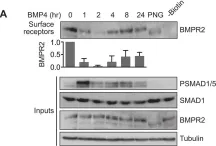
In Int J Mol Sci on 26 January 2023 by Song, Y. S., Zaitoun, I. S., et al.
Fig.2.A

-
WB
-
Collected and cropped from Int J Mol Sci by CiteAb, provided under a CC-BY license
Image 1 of 13
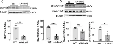
In Respir Res on 8 April 2022 by Kabwe, J. C., Sawada, H., et al.
Fig.1.C

-
WB
-
Collected and cropped from Respir Res by CiteAb, provided under a CC-BY license
Image 1 of 13
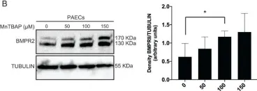
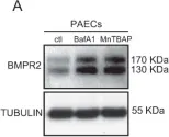
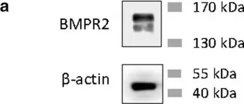
In Int J Mol Sci on 10 June 2020 by Gomez-Puerto, M. C., Sun, X. Q., et al.
Fig.1.B

-
WB
-
Collected and cropped from Int J Mol Sci by CiteAb, provided under a CC-BY license
Image 1 of 13
In Int J Mol Sci on 10 June 2020 by Gomez-Puerto, M. C., Sun, X. Q., et al.
Fig.1.A

-
WB
-
Collected and cropped from Int J Mol Sci by CiteAb, provided under a CC-BY license
Image 1 of 13
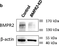
In Int J Mol Sci on 10 June 2020 by Gomez-Puerto, M. C., Sun, X. Q., et al.
Fig.3.B

-
WB
-
Collected and cropped from Int J Mol Sci by CiteAb, provided under a CC-BY license
Image 1 of 13
In Int J Mol Sci on 10 June 2020 by Gomez-Puerto, M. C., Sun, X. Q., et al.
Fig.3.A

-
WB
-
Collected and cropped from Int J Mol Sci by CiteAb, provided under a CC-BY license
Image 1 of 13
In Int J Mol Sci on 10 June 2020 by Gomez-Puerto, M. C., Sun, X. Q., et al.
Fig.3.C

-
WB
-
Collected and cropped from Int J Mol Sci by CiteAb, provided under a CC-BY license
Image 1 of 13
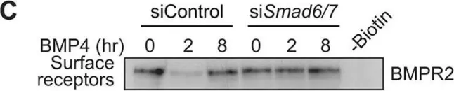
In J Cell Sci on 15 July 2019 by Miller, D. S. J., Schmierer, B., et al.
Fig.6.C

-
WB
-
Homo sapiens (Human)
Collected and cropped from J Cell Sci by CiteAb, provided under a CC-BY license
Image 1 of 13
In J Cell Sci on 15 July 2019 by Miller, D. S. J., Schmierer, B., et al.
Fig.5.A

-
WB
-
Homo sapiens (Human)
Collected and cropped from J Cell Sci by CiteAb, provided under a CC-BY license
Image 1 of 13
In BMC Res Notes on 11 June 2019 by Gorrell, R. E., Totten, M. H., et al.
Fig.2.A

-
WB
-
Homo sapiens (Human)
Collected and cropped from BMC Res Notes by CiteAb, provided under a CC-BY license
Image 1 of 13
In BMC Res Notes on 11 June 2019 by Gorrell, R. E., Totten, M. H., et al.
Fig.3.B

-
WB
-
Homo sapiens (Human)
Collected and cropped from BMC Res Notes by CiteAb, provided under a CC-BY license
Image 1 of 13
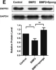
In PLoS One on 19 December 2013 by Sun, Q., Mao, S., et al.
Fig.2.E

-
WB
-
Collected and cropped from PLoS One by CiteAb, provided under a CC-BY license
Image 1 of 13
In PLoS One on 19 December 2013 by Sun, Q., Mao, S., et al.
Fig.3.E

-
WB
-
Collected and cropped from PLoS One by CiteAb, provided under a CC-BY license
Image 1 of 13