Ovarian cancer is the most lethal gynecological cancer, and it is frequently diagnosed at advanced stages, with recurrences after treatments. Treatment failure and resistance are due to hypoxia-inducible factors (HIFs) activated by cancer cells adapt to hypoxia. IGFBP3, which was previously identified as a growth/invasion/metastasis suppressor of ovarian cancer, plays a key role in inhibiting tumor angiogenesis. Although IGFBP3 can effectively downregulate tumor proliferation and vasculogenesis, its effects are only transient. Tumors enter a hypoxic state when they grow large and without blood vessels; then, the tumor cells activate HIFs to regulate cell metabolism, proliferation, and induce vasculogenesis to adapt to hypoxic stress. After IGFBP3 was transiently expressed in highly invasive ovarian cancer cell line and heterotransplant on mice, the xenograft tumors demonstrated a transient growth arrest with de-vascularization, causing tumor cell hypoxia. Tumor re-proliferation was associated with early HIF-1α and later HIF-2α activations. Both HIF-1α and HIF-2α were related to IGFBP3 expressions. In the down-expression of IGFBP3 in xenograft tumors and transfectants, HIF-2α was the major activated protein. This study suggests that HIF-2α presentation is crucial in the switching of epithelial ovarian cancer from dormancy to proliferation states. In highly invasive cells, the cancer hallmarks associated with aggressiveness could be activated to escape from the growth restriction state.
© 2021. The Author(s).
Applications
Reactivity
Research Area
In Scientific Reports on 25 November 2021 by Shih, H. J., Chang, H. F., et al.
-
WB
-
Cancer Research
In Cytokine on 1 January 2021 by Minuzzi, L. G., da Conceição, L. R., et al.
Interleukin-15 (IL-15) is a myokine that has been proposed to modulate skeletal muscle and adipose tissue mass, as well as insulin sensitivity. However, the evidence suggesting a role for IL-15 in improving whole-body insulin sensitivity and decreasing adiposity comes mainly from studies using supraphysiological levels of this cytokine. This study examined the effect of a short-term exercise training protocol on the protein content of IL-15, it's signaling pathway, and glucose tolerance in aged rats.
Fourteen Wistar rats were divided into Young Sedentary (Young, n = 4); Old Sedentary (Old, n = 5); Old Exercise (Old.Exe, n = 5) groups. The animals from the exercised group were submitted to a short-term physical exercise protocol for five days. At the end of physical training and after 16 h of the last exercise session, the animals were euthanized, and tissue collection was done.
Physical exercise decreased epididymal and mesenteric fat mass and promoted positive effects on glucose tolerance and insulin sensitivity. Muscle IL-15 protein levels were not changed following the short-term physical exercise training with no alterations in the post-exercise IL-15-JAK/STAT signaling pathway. We found a tendency to increased HIF1α and a significant increase in its regulator, PHD2, in the skeletal muscle after exercise.
The elderly rats submitted to short-term aerobic physical training did not present skeletal muscle alteration in the protein content of the IL-15 and IL-15-JAK/STAT signaling pathway. However, short-term aerobic physical training was able to modulate the expression of HIF1α and its regulator PHD2, suggesting an essential role of these proteins in improving post-exercise glucose tolerance and insulin sensitivity in elderly rats.
Copyright © 2020 Elsevier Ltd. All rights reserved.
-
WB
-
Rattus norvegicus (Rat)
-
Immunology and Microbiology
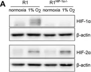
In Oncotarget on 13 October 2017 by Bino, L., Prochazkova, J., et al.
Fig.2.A

-
WB
-
Mus musculus (House mouse)
Collected and cropped from Oncotarget by CiteAb, provided under a CC-BY license
Image 1 of 2
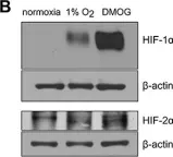
In Oncotarget on 13 October 2017 by Bino, L., Prochazkova, J., et al.
Fig.2.B

-
WB
-
Mus musculus (House mouse)
Collected and cropped from Oncotarget by CiteAb, provided under a CC-BY license
Image 1 of 2


